- Calcium & bone metabolism
- Persistence with Denosumab in Male Osteoporosis Patients: A Real-World, Non-Interventional Multicenter Study
-
Chaiho Jeong, Jeongmin Lee, Jinyoung Kim, Jeonghoon Ha, Kwanhoon Jo, Yejee Lim, Mee Kyoung Kim, Hyuk-Sang Kwon, Tae-Seo Sohn, Ki-Ho Song, Moo Il Kang, Ki-Hyun Baek
-
Endocrinol Metab. 2023;38(2):260-268. Published online April 27, 2023
-
DOI: https://doi.org/10.3803/EnM.2023.1663
-
-
1,798
View
-
111
Download
-
1
Crossref
-
 Abstract Abstract
 PDF PDF Supplementary Material Supplementary Material PubReader PubReader  ePub ePub
- Background
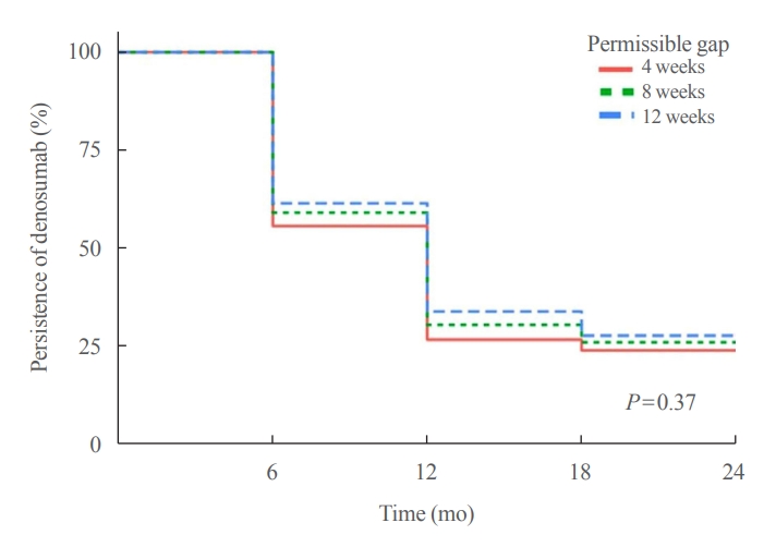
Persistence with denosumab in male patients has not been adequately investigated, although poor denosumab persistence is associated with a significant risk of rebound vertebral fractures.
Methods
We retrospectively evaluated 294 Korean male osteoporosis patients treated with denosumab at three medical centers and examined their persistence with four doses of denosumab injection over 24 months of treatment. Persistence was defined as the extent to which a patient adhered to denosumab treatment in terms of the prescribed interval and dose, with a permissible gap of 8 weeks. For patients who missed their scheduled treatment appointment(s) during the follow-up period (i.e., no-shows), Cox proportional regression analysis was conducted to explore the factors associated with poor adherence. Several factors were considered, such as age, prior anti-osteoporotic drug use, the treatment provider’s medical specialty, the proximity to the medical center, and financial burdens of treatment.
Results
Out of 294 male patients, 77 (26.2%) completed all four sequential rounds of the denosumab treatment. Out of 217 patients who did not complete the denosumab treatment, 138 (63.6%) missed the scheduled treatment(s). Missing treatment was significantly associated with age (odds ratio [OR], 1.03), prior bisphosphonate use (OR, 0.76), and prescription by non-endocrinologists (OR, 2.24). Denosumab was stopped in 44 (20.3%) patients due to medical errors, in 24 (11.1%) patients due to a T-score improvement over –2.5, and in five (2.3%) patients due to expected dental procedures.
Conclusion
Our study showed that only one-fourth of Korean male osteoporosis patients were fully adherent to 24 months of denosumab treatment.
-
Citations
Citations to this article as recorded by  - Denosumab
Reactions Weekly.2023; 1963(1): 206. CrossRef
- Thyroid
- Usefulness of Real-Time Quantitative Microvascular Ultrasonography for Differentiation of Graves’ Disease from Destructive Thyroiditis in Thyrotoxic Patients
-
Han-Sang Baek, Ji-Yeon Park, Chai-Ho Jeong, Jeonghoon Ha, Moo Il Kang, Dong-Jun Lim
-
Endocrinol Metab. 2022;37(2):323-332. Published online April 13, 2022
-
DOI: https://doi.org/10.3803/EnM.2022.1413
-
-
3,723
View
-
144
Download
-
5
Web of Science
-
5
Crossref
-
Abstract
PDFSupplementary MaterialPubReader ePub
- Background
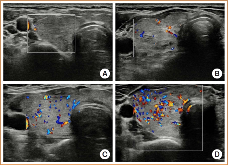
Microvascular ultrasonography (MVUS) is a third-generation Doppler technique that was developed to increase sensitivity compared to conventional Doppler. The purpose of this study was to compare MVUS with conventional color Doppler (CD) and power Doppler (PD) imaging to distinguish Graves’ disease (GD) from destructive thyroiditis (DT).
Methods
This prospective study included 101 subjects (46 GDs, 47 DTs, and eight normal controls) from October 2020 to November 2021. All ultrasonography examinations were performed using microvascular flow technology (MV-Flow). The CD, PD, and MVUS images were semi-quantitatively graded according to blood flow patterns. On the MVUS images, vascularity indices (VIs), which were the ratio (%) of color pixels in the total grayscale pixels in a defined region of interest, were obtained automatically. Receiver operating characteristic curve analysis was performed to verify the diagnostic performance of MVUS. The interclass correlation coefficient and Cohen’s kappa analysis were used to analyze the reliability of MVUS (ClinicalTrials.gov:NCT04879173).
Results
The area under the curve (AUC) for CD, PD, MVUS, and MVUS-VI was 0.822, 0.844, 0.808, and 0.852 respectively. The optimal cutoff value of the MVUS-VI was 24.95% for distinguishing GD and DT with 87% sensitivity and 80.9% specificity. We found a significant positive correlation of MVUS-VI with thyrotropin receptor antibody (r=0.554) and with thyroid stimulating immunoglobulin bioassay (r=0.841). MVUS showed high intra- and inter-observer reliability from various statistical method.
Conclusion
In a real time and quantitative manner, MVUS-VI could be helpful to differentiate GD from thyroiditis in thyrotoxic patients, with less inter-observer variability.
-
Citations
Citations to this article as recorded by - Association of autoimmune thyroid disease with type 1 diabetes mellitus and its ultrasonic diagnosis and management
Jin Wang, Ke Wan, Xin Chang, Rui-Feng Mao
World Journal of Diabetes.2024; 15(3): 348. CrossRef - The Early Changes in Thyroid-Stimulating Immunoglobulin Bioassay over Anti-Thyroid Drug Treatment Could Predict Prognosis of Graves’ Disease
Jin Yu, Han-Sang Baek, Chaiho Jeong, Kwanhoon Jo, Jeongmin Lee, Jeonghoon Ha, Min Hee Kim, Jungmin Lee, Dong-Jun Lim
Endocrinology and Metabolism.2023; 38(3): 338. CrossRef - Duplex Hemodynamic Parameters of Both Superior and Inferior Thyroid Arteries in Evaluation of Thyroid Hyperfunction Disorders
Maha Assem Hussein, Alaa Abdel Hamid, Rasha M Abdel Samie, Elshaymaa Hussein, Shereen Sadik Elsawy
International Journal of General Medicine.2022; Volume 15: 7131. CrossRef - Case 5: A 41-Year-Old Woman With Palpitation
Jiwon Yang, Kabsoo Shin, Jeongmin Lee, Jeonghoon Ha, Dong-Jun Lim, Han-Sang Baek
Journal of Korean Medical Science.2022;[Epub] CrossRef - Microvascular assessment of fascio-cutaneous flaps by ultrasound: A large animal study
Guillaume Goudot, Yanis Berkane, Eloi de Clermont-Tonnerre, Claire Guinier, Irina Filz von Reiterdank, Antonia van Kampen, Korkut Uygun, Curtis L. Cetrulo, Basak E. Uygun, Anahita Dua, Alexandre G. Lellouch
Frontiers in Physiology.2022;[Epub] CrossRef
- Calcium & Bone Metabolism
- Changes in Serum Dickkopf-1, RANK Ligand, Osteoprotegerin, and Bone Mineral Density after Allogeneic Hematopoietic Stem Cell Transplantation Treatment
-
Eunhee Jang, Jeonghoon Ha, Ki-Hyun Baek, Moo Il Kang
-
Endocrinol Metab. 2021;36(6):1211-1218. Published online December 8, 2021
-
DOI: https://doi.org/10.3803/EnM.2021.1248
-
-
3,023
View
-
104
Download
-
1
Web of Science
-
2
Crossref
-
Abstract
PDFPubReader ePub
- Background
Dickkopf-1 (DKK1) regulates bone formation by inhibiting canonical Wnt/β-catenin pathway signaling, and indirectly enhances osteoclastic activity by altering the expression ratio of receptor activator of nuclear factor-κB ligand (RANKL) relative to osteoprotegerin (OPG). However, it is difficult to explain continued bone loss after allogeneic stem cell transplantation (allo-SCT) in terms of changes in only RANKL and OPG. Few studies have evaluated changes in DKK1 after allo-SCT.
Methods
We prospectively enrolled 36 patients with hematologic malignancies who were scheduled for allo-SCT treatment. Serum DKK1, OPG, and RANKL levels were measured before (baseline), and at 1, 4, 12, 24, and 48 weeks after allo-SCT treatment. Bone mineral density (BMD) was assessed using dual-energy X-ray absorptiometry before (baseline) and 24 and 48 weeks after allo-SCT treatment.
Results
After allo-SCT treatment, the DKK1 level decreased rapidly, returned to baseline during the first 4 weeks, and remained elevated for 48 weeks (P<0.0001 for changes observed over time). The serum RANKL/OPG ratio peaked at 4 weeks and then declined (P<0.001 for changes observed over time). BMD decreased relative to the baseline at all timepoints during the study period, and the lumbar spine in female patients had the largest decline (–11.3%±1.6% relative to the baseline at 48 weeks, P<0.05).
Conclusion
Serum DKK1 levels rapidly decreased at 1 week and then continued to increase for 48 weeks; bone mass decreased for 48 weeks following engraftment in patients treated with allo-SCT, suggesting that DKK1-mediated inhibition of osteoblast differentiation plays a role in bone loss in patients undergoing allo-SCT.
-
Citations
Citations to this article as recorded by - Fracture risk and assessment in adults with cancer
Carrie Ye, William D. Leslie
Osteoporosis International.2023; 34(3): 449. CrossRef - Short-Term Impact of Hematopoietic Stem Cell Transplantation in Leukemia Patients on Bone Bio Markers, Electrolytes and Blood Profile
Rhythm Joshi, Zehva Khan, Aakriti Garg, Dinesh Bhurani, Nidhi B Agarwal, Ubada Aqeel, Mohd Ashif Khan
OBM Transplantation.2023; 07(02): 1. CrossRef
- Bone Metabolism
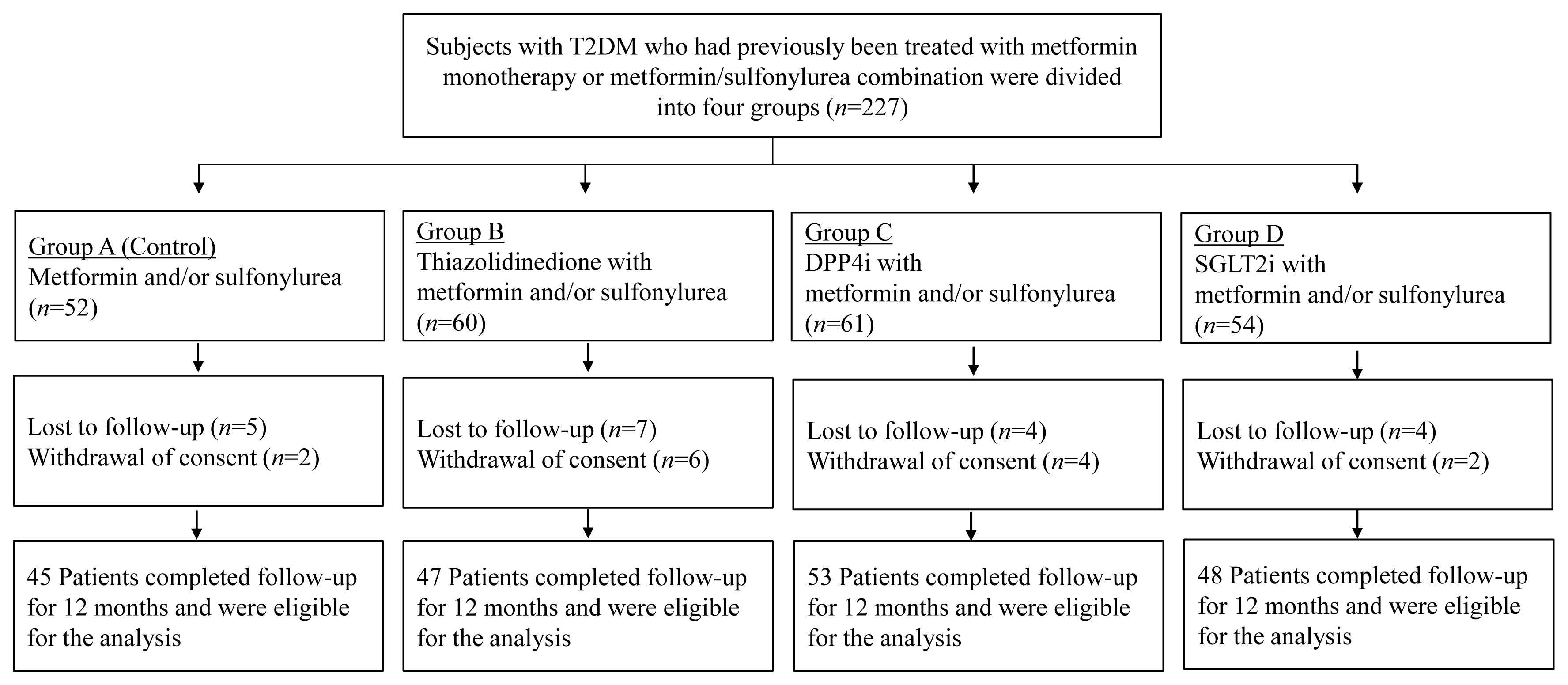
- Comparison of the Effects of Various Antidiabetic Medication on Bone Mineral Density in Patients with Type 2 Diabetes Mellitus
-
Jeonghoon Ha, Yejee Lim, Mee Kyoung Kim, Hyuk-Sang Kwon, Ki-Ho Song, Seung Hyun Ko, Moo Il Kang, Sung Dae Moon, Ki-Hyun Baek
-
Endocrinol Metab. 2021;36(4):895-903. Published online August 9, 2021
-
DOI: https://doi.org/10.3803/EnM.2021.1026
-
-
6,173
View
-
230
Download
-
4
Web of Science
-
4
Crossref
-
Abstract
PDFPubReader ePub
- Background
Prospective comparative studies on the effects of various antidiabetic agents on bone metabolism are limited. This study aimed to assess changes in bone mass and biochemical bone markers in postmenopausal patients with type 2 diabetes mellitus (T2DM).
Methods
This prospective, multicenter, open-label, comparative trial included 264 patients with T2DM. Patients who had received a metformin, or sulfonylurea/metformin combination (Group 1); a thiazolidinedione combination (Group 2); a dipeptidyl peptidase-4 inhibitor (gemigliptin) combination (Group 3); or an sodium-glucose cotransporter 2 inhibitor (empagliflozin) combination (Group 4) were prospectively treated for 12 months; bone mineral density (BMD) and bone turnover marker (BTM) changes were evaluated.
Results
The femoral neck BMD percentage changes were −0.79%±2.86% (Group 1), −2.50%±3.08% (Group 2), −1.05%±2.74% (Group 3), and −1.24%±2.91% (Group 4) (P<0.05). The total hip BMD percentage changes were −0.57%±1.79% (Group 1), −1.74%±1.48% (Group 2), −0.75%±1.87% (Group 3), and −1.27%±1.72% (Group 4) (P<0.05). Mean serum BTM (C-terminal type 1 collagen telopeptide and procollagen type 1 amino-terminal propeptide) levels measured during the study period did not change over time or differ between groups.
Conclusion
Significant bone loss in the femoral neck and total hip was associated with thiazolidinedione combination regimens. However, bone loss was not significantly associated with combination regimens including gemigliptin or empagliflozin. Caution should be exercised during treatment with antidiabetic medications that adversely affect the bone in patients with diabetes at a high risk of bone loss.
-
Citations
Citations to this article as recorded by - Meta-Analysis on the Association Between DPP-4 Inhibitors and Bone Mineral Density and Osteoporosis
Lili Huang, Wei Zhong, Xinghuan Liang, Huijuan Wang, Shi-en Fu, Zuojie Luo
Journal of Clinical Densitometry.2024; 27(1): 101455. CrossRef - A multicentre, double‐blind, placebo‐controlled, randomized, parallel comparison, phase 3 trial to evaluate the efficacy and safety of pioglitazone add‐on therapy in type 2 diabetic patients treated with metformin and dapagliflozin
Soo Lim, Seung‐Hwan Lee, Kyung‐Wan Min, Chang Beom Lee, Sang Yong Kim, Hye Jin Yoo, Nan Hee Kim, Jae Hyeon Kim, Seungjoon Oh, Jong Chul Won, Hyuk Sang Kwon, Mi Kyung Kim, Jung Hwan Park, In‐Kyung Jeong, Sungrae Kim
Diabetes, Obesity and Metabolism.2024;[Epub] CrossRef - Association of Bone Turnover Markers with Type 2 Diabetes Mellitus and Microvascular Complications: A Matched Case-Control Study
Yilin Hou, Xiaoyu Hou, Qian Nie, Qiuyang Xia, Rui Hu, Xiaoyue Yang, Guangyao Song, Luping Ren
Diabetes, Metabolic Syndrome and Obesity.2023; Volume 16: 1177. CrossRef - Complementary effects of dapagliflozin and lobeglitazone on metabolism in a diet-induced obese mouse model
Yun Kyung Lee, Tae Jung Oh, Ji In Lee, Bo Yoon Choi, Hyen Chung Cho, Hak Chul Jang, Sung Hee Choi
European Journal of Pharmacology.2023; 957: 175946. CrossRef
- Hypothalamus and Pituitary Gland
- Heart Rate Variability in Postoperative Patients with Nonfunctioning Pituitary Adenoma
-
Jeonghoon Ha, Hansang Baek, Chaiho Jeong, Minsoo Yeo, Seung-Hwan Lee, Jae Hyoung Cho, Ki-Hyun Baek, Moo Il Kang, Dong-Jun Lim
-
Endocrinol Metab. 2021;36(3):678-687. Published online June 10, 2021
-
DOI: https://doi.org/10.3803/EnM.2021.978
-
-
4,591
View
-
109
Download
-
3
Web of Science
-
3
Crossref
-
Abstract
PDFSupplementary MaterialPubReader ePub
- Background
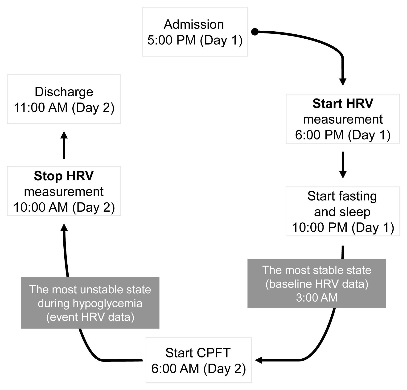
Decreased heart rate variability (HRV) has been reported to be associated with cardiac autonomic dysfunction. Hypopituitarism in nonfunctioning pituitary adenoma (NFPA) is often linked to increased cardiovascular mortality. We therefore hypothesized that postoperative NFPA patients with hormone deficiency have an elevated risk of HRV alterations indicating cardiac autonomic dysfunction.
Methods
A total of 22 patients with NFPA were enrolled in the study. Between 3 and 6 months after surgery, a combined pituitary function test (CPFT) was performed, and HRV was measured. The period of sleep before the CPFT was deemed the most stable period, and the hypoglycemic period that occurred during the CPFT was defined as the most unstable period. Changes in HRV parameters in stable and unstable periods were observed and compared depending on the status of hormone deficiencies.
Results
In patients with adrenocorticotropic hormone (ACTH) deficiency with other pituitary hormone deficiencies, the low frequency to high frequency ratio, which represents overall autonomic function and is increased in the disease state, was higher (P=0.005). Additionally, the standard deviation of the normal-to-normal interval, which decreases in the autonomic dysfunction state, was lower (P=0.030) during the hypoglycemic period. In panhypopituitarism, the low frequency to high frequency ratio during the hypoglycemic period was increased (P=0.007).
Conclusion
HRV analysis during CPFT enables estimation of cardiac autonomic dysfunction in patients with NFPA who develop ACTH deficiency with other pituitary hormone deficiencies or panhypopituitarism after surgery. These patients may require a preemptive assessment of cardiovascular risk.
-
Citations
Citations to this article as recorded by - Heart Rate Variability in Subjects with Severe Allergic Background Undergoing COVID-19 Vaccination
Maria Bernadette Cilona, Filippo D’Amico, Chiara Asperti, Giuseppe Alvise Ramirez, Stefano Turi, Giovanni Benanti, Shai Marc Bohane, Serena Nannipieri, Rosa Labanca, Matteo Gervasini, Federica Russetti, Naomi Viapiana, Martina Lezzi, Giovanni Landoni, Lor
Vaccines.2023; 11(3): 567. CrossRef - Pituitary Diseases and COVID-19 Outcomes in South Korea: A Nationwide Cohort Study
Jeonghoon Ha, Kyoung Min Kim, Dong-Jun Lim, Keeho Song, Gi Hyeon Seo
Journal of Clinical Medicine.2023; 12(14): 4799. CrossRef - Effect of a 16-Session Qigong Program in Non-Hodgkin Lymphoma Survivors: A Randomized Clinical Trial
Keyla Vargas-Román, Emilia I. De la Fuente-Solana, Jonathan Cortés-Martín, Juan Carlos Sánchez-García, Christian J. González-Vargas, Lourdes Díaz-Rodríguez
Journal of Clinical Medicine.2022; 11(12): 3421. CrossRef
- Clinical Study
- Association of Hyperparathyroidism and Papillary Thyroid Cancer: A Multicenter Retrospective Study
-
Chaiho Jeong, Hye In Kwon, Hansang Baek, Hun-Sung Kim, Dong-Jun Lim, Ki-Hyun Baek, Jeonghoon Ha, Moo Il Kang
-
Endocrinol Metab. 2020;35(4):925-932. Published online December 10, 2020
-
DOI: https://doi.org/10.3803/EnM.2020.725
-
-
5,396
View
-
184
Download
-
9
Web of Science
-
8
Crossref
-
Abstract
PDFPubReader ePub
- Background
Concomitant papillary thyroid cancer (PTC) and hyperparathyroidism (HPT) have been reported in several studies. Our study aimed to investigate the incidence of concomitant PTC in HPT patients upon preoperative diagnosis and present a clinical opinion on detecting thyroid malignancy in case of parathyroidectomy.
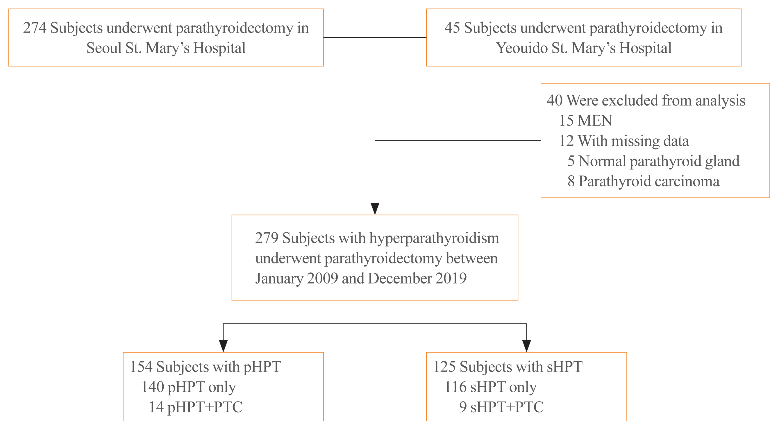
Methods
Patients who underwent parathyroidectomy between January 2009 and December 2019 in two medical centers were included. Of the 279 participants 154 were diagnosed as primary hyperparathyroidism (pHPT) and 125 as secondary hyperparathyroidism (sHPT). The incidence of concomitant PTC and its clinical characteristics were compared with 98 patients who underwent thyroidectomy and were diagnosed with classical PTC during the same period.
Results
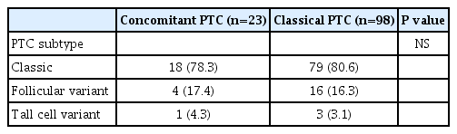
Concurrent PTC was detected in 14 patients (9.1%) with pHPT and in nine patients (7.2%) with sHPT. Ten (71.4%) and seven (77.8%) PTCs were microcarcinomas in the pHPT and sHPT cases respectively. In the pHPT patients, vitamin D was lower in the pHPT+PTC group (13.0±3.7 ng/mL) than in the pHPT-only group (18.5±10.4 ng/mL; P=0.01). Vitamin D levels were also lower in the sHPT+PTC group (12.3±5.6 ng/mL) than in the sHPT-only group (18.0±10.2 ng/mL; P=0.12). In the concomitant PTC group, lymph node ratio was higher than in the classical PTC group (P=0.00).
Conclusion
A high prevalence of concomitant PTC was seen in patients with pHPT and sHPT. Those concomitant PTCs were mostly microcarcinomas and had more aggressive features, suggesting that efforts should be made to detect concomitant malignancies in the preoperative parathyroidectomy evaluation.
-
Citations
Citations to this article as recorded by - The unexpected effect of parathyroid adenoma on inflammation
Ahmet Tarik Harmantepe, Belma Kocer, Zulfu Bayhan, Emre Gonullu, Ugur Can Dulger
Updates in Surgery.2024; 76(2): 589. CrossRef - Evaluation of Nodular Goiter and Papillary Thyroid Cancer Coincidence in Patients with Primary Hyperparathyroidism
Mustafa ÇALIŞKAN, Hasret CENGİZ, Taner DEMİRCİ
Düzce Tıp Fakültesi Dergisi.2023; 25(2): 200. CrossRef - Papillary thyroid carcinoma coexisting with benign thyroid and parathyroid pathology: clinical and pathomorphological features
A. Dinets, M. Gorobeiko, V. Hoperia, A. Lovin, S. Tarasenko
INTERNATIONAL JOURNAL OF ENDOCRINOLOGY (Ukraine).2023; 19(4): 274. CrossRef - The Nexus of Hyperparathyroidism and Thyroid Carcinoma: Insights into Pathogenesis and Diagnostic Challenges—A Narrative Review
Gregorio Scerrino, Nunzia Cinzia Paladino, Giuseppina Orlando, Giuseppe Salamone, Pierina Richiusa, Stefano Radellini, Giuseppina Melfa, Giuseppa Graceffa
Journal of Clinical Medicine.2023; 13(1): 147. CrossRef - Is preoperative parathyroid localization necessary for tertiary hyperparathyroidism?
Rongzhi Wang, Peter Abraham, Brenessa Lindeman, Herbert Chen, Jessica Fazendin
The American Journal of Surgery.2022; 224(3): 918. CrossRef - Papillary thyroid carcinoma prevalence and its predictors in patients with primary hyperparathyroidism
Elif Tutku DURMUŞ, Ayşegül ATMACA, Mehmet KEFELİ, Ramis ÇOLAK, Buğra DURMUŞ, Cafer POLAT
Journal of Health Sciences and Medicine.2022; 5(5): 1499. CrossRef - Association of Hyperparathyroidism and Papillary Thyroid Cancer: A Multicenter Retrospective Study (Endocrinol Metab 2020;35:925-32, Chaiho Jeong et al.)
Chaiho Jeong, Jeonghoon Ha, Moo Il Kang
Endocrinology and Metabolism.2021; 36(1): 205. CrossRef - Association of Hyperparathyroidism and Papillary Thyroid Cancer: A Multicenter Retrospective Study (Endocrinol Metab 2020;35:925-32, Chaiho Jeong et al.)
Burcu Candemir, Coşkun Meriç
Endocrinology and Metabolism.2021; 36(1): 203. CrossRef
- Clinical Study
- Comparison of Natural Course between Thyroid Cancer Nodules and Thyroid Benign Nodules
-
Kyun-Jin Yun, Jeonghoon Ha, Min-Hee Kim, Ye Young Seo, Mee Kyoung Kim, Hyuk-Sang Kwon, Ki-Ho Song, Moo Il Kang, Ki-Hyun Baek
-
Endocrinol Metab. 2019;34(2):195-202. Published online June 24, 2019
-
DOI: https://doi.org/10.3803/EnM.2019.34.2.195
-
-
4,536
View
-
65
Download
-
8
Web of Science
-
8
Crossref
-
Abstract
PDFPubReader ePub
- Background
The natural course of thyroid cancer nodules and benign nodules is different. This study was to compare the changes in size between thyroid cancer nodules and thyroid benign nodules. The risk factors associated with the changes of thyroid cancer nodules were assessed. MethodsThis study contains retrospective observational and prospective analysis. A total of 113 patients with 120 nodules were recruited in the cancer group, and 116 patients with 119 nodules were enrolled in the benign group. Thyroid ultrasonography was performed at least two times at more than 1-year interval. ResultsThe mean follow-up durations were 29.5±18.8 months (cancer group) and 31.9±15.8 months (benign group) (P=0.32). The maximum diameter change in length was 0.36±0.97 mm/year in the cancer group and –0.04±0.77 mm/year in the benign group (P<0.01). The volume was significantly increased in the cancer group compared with the benign group (0.06±0.18 mL/year vs. 0.004±0.05 mL/year, respectively, P<0.01; 26.9%±57.9%/year vs. 1.7%±26.0%/year, P<0.01). Initial maximum diameter (β=0.02, P<0.01) and initial volume (β=0.13, P<0.01) were significantly associated with volume change (mL)/year. Initial maximum standardized uptake value did not predict the nodule growth. ConclusionIt is suggested that thyroid cancer nodules progress rapidly compared with benign nodules. Initial size and volume of nodule were independent risk factors for cancer nodule growth.
-
Citations
Citations to this article as recorded by - RAS-Mutated Cytologically Indeterminate Thyroid Nodules: Prevalence of Malignancy and Behavior Under Active Surveillance
Hannah J. Sfreddo, Elizabeth S. Koh, Karena Zhao, Christina E. Swartzwelder, Brian R. Untch, Jennifer L. Marti, Benjamin R. Roman, Jared Dublin, Ronald S. Wang, Rong Xia, Jean-Marc Cohen, Bin Xu, Ronald Ghossein, Babak Givi, Jay O. Boyle, R. Michael Tuttl
Thyroid®.2024;[Epub] CrossRef - Ultrasound for the assessment of thyroid nodules: an overview for non-radiologists
Conor Hamill, Peter Ellis, Philip C Johnston
British Journal of Hospital Medicine.2022; 83(7): 1. CrossRef - Цитологічно підтверджений вузловий зоб у членів Українсько-Американського когортного дослідження: дескриптивний аналіз результатів обстеження за 1998- 2015 роки
M.D. Tronko, L.S. Strafun, H.M. Terekhova, H.A. Zamotayeva, I.P. Pasteur
Endokrynologia.2022; 27(1): 5. CrossRef - A Computational Study on the Role of Parameters for Identification of Thyroid Nodules by Infrared Images (and Comparison with Real Data)
José R. González, Charbel Damião, Maira Moran, Cristina A. Pantaleão, Rubens A. Cruz, Giovanna A. Balarini, Aura Conci
Sensors.2021; 21(13): 4459. CrossRef - Ultrasound in active surveillance for low-risk papillary thyroid cancer: imaging considerations in case selection and disease surveillance
Sangeet Ghai, Ciara O’Brien, David P. Goldstein, Anna M. Sawka, Lorne Rotstein, Dale Brown, John de Almeida, Patrick Gullane, Ralph Gilbert, Douglas Chepeha, Jonathan Irish, Jesse Pasternak, Shereen Ezzat, James P. Brierley, Richard W. Tsang, Eric Monteir
Insights into Imaging.2021;[Epub] CrossRef - Association between various thyroid gland diseases, TSH values and thyroid cancer: a case–control study
Leif Schiffmann, Karel Kostev, Matthias Kalder
Journal of Cancer Research and Clinical Oncology.2020; 146(11): 2989. CrossRef - Combination of peroxisome proliferator–activated receptor gamma and retinoid X receptor agonists induces sodium/iodide symporter expression and inhibits cell growth of human thyroid cancer cells
Jui-Yu Chen, Jane-Jen Wang, Hsin-Chen Lee, Chin-Wen Chi, Chen-Hsen Lee, Yi-Chiung Hsu
Journal of the Chinese Medical Association.2020; 83(10): 923. CrossRef - Growth rates of malignant and benign thyroid nodules in an ultrasound follow-up study: a retrospective cohort study
Michael Cordes, Theresa Ida Götz, Karen Horstrup, Torsten Kuwert, Christian Schmidkonz
BMC Cancer.2019;[Epub] CrossRef
- Thyroid
- Natural Course of Cytologically Benign Thyroid Nodules: Observation of Ultrasonographic Changes
-
Dong Jun Lim, Jee Young Kim, Ki Hyun Baek, Mee Kyoung Kim, Woo Chan Park, Jong Min Lee, Moo Il Kang, Bong Yun Cha
-
Endocrinol Metab. 2013;28(2):110-118. Published online June 18, 2013
-
DOI: https://doi.org/10.3803/EnM.2013.28.2.110
-
-
4,311
View
-
37
Download
-
18
Web of Science
-
23
Crossref
-
Abstract
PDFPubReader
- Background
The natural course of cytologically benign thyroid nodules remains unclear. The aim of this study was to evaluate whether ultrasonographic (US) changes are associated with changes in nodule volume during follow-up. MethodsWe retrospectively reviewed over 4 years of clinical records of patients with benign thyroid nodules as confirmed by fine needle aspiration (FNA). In total, 186 patients with 202 benign thyroid nodules were included for study. We assessed for changes in nodule volume and examined the cystic portion of the nodule as well as four US features (echogenicity, margin, calcification pattern, and shape). ResultsDuring follow-up (mean, 21.7±10.7 months) and using 50% as a cutoff value, nodule volumes increased in 11.8%, exhibited no change in 79.9%, and decreased in 8.3% of patients. Proportion of nodules demonstrating at least one US change was 20.8% (42/202). The most common US changes (in descending order of frequency) were cystic change, margin change, and calcification pattern change. Nodule shape and echogenicity rarely changed. Increased nodule volume was not significantly associated with any US features or with the number of FNAs but was associated with younger age at time of diagnosis. ConclusionAlthough a portion of thyroid nodules confirmed as benign showed US changes or volume changes during the follow-up period, these findings may only represent the natural course of benign nodules. Frequent follow-up with US might be needed for only a small number of cases with suspicious US findings.
-
Citations
Citations to this article as recorded by - Follow-up of benign thyroid nodules confirmed by ultrasound-guided core needle biopsy after inconclusive cytology on fine-needle aspiration biopsy
Yoon Ji Hwang, Hye Ryoung Koo, Jeong Seon Park
Ultrasonography.2023; 42(1): 121. CrossRef - Clinical Characteristics, Diagnostic Approach and Outcome of Thyroid Incidental Findings vs. Clinically Overt Thyroid Nodules: An Observational Single-Centre Study
Tom Jansen, Nike Stikkelbroeck, Annenienke van de Ven, Ilse van Engen-van Grunsven, Marcel Janssen, Han Bonenkamp, Martin Gotthardt, Romana T. Netea-Maier
Cancers.2023; 15(8): 2350. CrossRef - Association between various thyroid gland diseases, TSH values and thyroid cancer: a case–control study
Leif Schiffmann, Karel Kostev, Matthias Kalder
Journal of Cancer Research and Clinical Oncology.2020; 146(11): 2989. CrossRef - TI-RADS und andere sonografische Klassifikationssysteme für Schilddrüsenknoten
Julian M. M. Rogasch, Christoph Wetz, Winfried Brenner
Onkologie up2date.2020; 2(03): 223. CrossRef - TI-RADS und andere sonografische Klassifikationssysteme für Schilddrüsenknoten
Julian M.M. Rogasch, Christoph Wetz, Winfried Brenner
Radiopraxis.2020; 13(01): E1. CrossRef - Changes of Nodular Size and Its Risk Factors in Iodine-Sufficient Area: a Retrospective Cohort Analysis of 7753 Thyroid Nodules
Hwa Young Ahn, Kyung Won Kim, Hoon Sung Choi, Jae Hoon Moon, Ka Hee Yi, Min Kyung Hyun, Min Joo Kang, Jung Im Shim, Ja Youn Lee, Do Joon Park, Young Joo Park
International Journal of Thyroidology.2020; 13(2): 118. CrossRef - Comparison of Natural Course between Thyroid Cancer Nodules and Thyroid Benign Nodules
Kyun-Jin Yun, Jeonghoon Ha, Min-Hee Kim, Ye Young Seo, Mee Kyoung Kim, Hyuk-Sang Kwon, Ki-Ho Song, Moo Il Kang, Ki-Hyun Baek
Endocrinology and Metabolism.2019; 34(2): 195. CrossRef - Risk factors for hypothyroidism in euthyroid thyroid nodule patients with lymphocytic thyroiditis on fine needle aspiration cytology
Jeong-Min Lee, Jeonghoon Ha, Kwanhoon Jo, Yejee Lim, Min-Hee Kim, Chan-Kwan Jung, So-Lyung Jung, Moo-Il Kang, Bong-Yun Cha, Dong-Jun Lim
The Korean Journal of Internal Medicine.2019; 34(6): 1287. CrossRef - Evaluation of the natural course of thyroid nodules in patients with acromegaly
Sema Ciftci Dogansen, Artur Salmaslioglu, Gulsah Yenidunya Yalin, Seher Tanrikulu, Sema Yarman
Pituitary.2019; 22(1): 29. CrossRef - TI-RADS und andere sonografische Klassifikationssystemefür Schilddrüsenknoten
Julian M.M. Rogasch, Christoph Wetz, Winfried Brenner
Der Nuklearmediziner.2019; 42(03): 206. CrossRef - Molecular profiling of thyroid nodule fine-needle aspiration cytology
Markus Eszlinger, Lorraine Lau, Sana Ghaznavi, Christopher Symonds, Shamir P. Chandarana, Moosa Khalil, Ralf Paschke
Nature Reviews Endocrinology.2017; 13(7): 415. CrossRef - Diagnostic accuracy of thyroid nodule growth to predict malignancy in thyroid nodules with benign cytology: systematic review and meta‐analysis
Naykky Singh Ospina, Spyridoula Maraka, Ana Espinosa DeYcaza, Derek O'Keeffe, Juan P. Brito, Michael R. Gionfriddo, M. Regina Castro, John C. Morris, Patricia Erwin, Victor M. Montori
Clinical Endocrinology.2016; 85(1): 122. CrossRef - Rapid thyroid nodule growth is not a marker for well-differentiated thyroid cancer
Claudius Falch, Steffen Axt, Bettina Scuffi, Alfred Koenigsrainer, Andreas Kirschniak, Sven Muller
World Journal of Surgical Oncology.2015;[Epub] CrossRef - Predicting the Size of Benign Thyroid Nodules and Analysis of Associated Factors That Affect Nodule Size
Seok Ho Seo, Tae Hyun Kim, Soon Ho Kim, Seung Hyun Lee, Jong Taek Kim, Dae Won Park, Dong Chul Lee
Chonnam Medical Journal.2015; 51(2): 97. CrossRef - Thyroid ultrasound findings in a follow-up survey of children from three Japanese prefectures: Aomori, Yamanashi and Nagasaki
Naomi Hayashida, Misa Imaizumi, Hiroki Shimura, Fumihiko Furuya, Noriyuki Okubo, Yasushi Asari, Takeshi Nigawara, Sanae Midorikawa, Kazuhiko Kotani, Shigeyuki Nakaji, Akira Ohtsuru, Takashi Akamizu, Masafumi Kitaoka, Shinichi Suzuki, Nobuyuki Taniguchi, S
Scientific Reports.2015;[Epub] CrossRef - Brief Review of Articles in 'Endocrinology and Metabolism' in 2013
Won-Young Lee
Endocrinology and Metabolism.2014; 29(3): 251. CrossRef - Natural Course of Cytologically Diagnosed Benign Thyroid Nodules
Eun-Kyung Kim
Journal of Korean Thyroid Association.2014; 7(2): 136. CrossRef - Ruling in or ruling out thyroid malignancy by molecular diagnostics of thyroid nodules
Markus Eszlinger, László Hegedüs, Ralf Paschke
Best Practice & Research Clinical Endocrinology & Metabolism.2014; 28(4): 545. CrossRef - Insufficient Experience in Thyroid Fine-Needle Aspiration Leads to Misdiagnosis of Thyroid Cancer
Jung Il Son, Sang Youl Rhee, Jeong-taek Woo, Won Seo Park, Jong Kyu Byun, Yu-Jin Kim, Ja Min Byun, Sang Ouk Chin, Suk Chon, Seungjoon Oh, Sung Woon Kim, Young Seol Kim
Endocrinology and Metabolism.2014; 29(3): 293. CrossRef - Clinical Outcomes in Patients with Non-Diagnostic Thyroid Fine Needle Aspiration Cytology: Usefulness of the Thyroid Core Needle Biopsy
Sung Hak Lee, Min Hee Kim, Ja Seong Bae, Dong Jun Lim, So Lyung Jung, Chan Kwon Jung
Annals of Surgical Oncology.2014; 21(6): 1870. CrossRef - Letter: Natural Course of Cytologically Benign Thyroid Nodules: Observation of Ultrasonographic Changes (Endocrinol Metab 2013;28:110-8, Dong Jun Lim et al.)
Sun Wook Cho
Endocrinology and Metabolism.2013; 28(3): 241. CrossRef - Natural Course of Benign Thyroid Nodules
Kyung Won Kim
Endocrinology and Metabolism.2013; 28(2): 94. CrossRef - Response: Natural Course of Cytologically Benign Thyroid Nodules: Observation of Ultrasonographic Changes (Endocrinol Metab 2013;28:110-8, Dong Jun Lim et al.)
Dong Jun Lim, Ki Hyun Baek
Endocrinology and Metabolism.2013; 28(3): 243. CrossRef
- Pregnancy-induced Osteoporosis Combined with Multiple Compression Fractures: A Case Report.
-
Ji Eun Lee, Jin Sun Jang, Sun Hee Ko, Min Hee Kim, Dong Jun Lim, Moo Il Kang, Bong Yun Cha, Sook Hee Hong, Ja seong Bae, Kyeoung Sik Ryu
-
Endocrinol Metab. 2011;26(2):150-154. Published online June 1, 2011
-
DOI: https://doi.org/10.3803/EnM.2011.26.2.150
-
-
2,093
View
-
28
Download
-
1
Crossref
-
Abstract
PDF
- Pregnancy associated osteoporosis (PAO) is a rare condition. It may affect women during pregnancy or after the delivery and it can induce severe back pain. Physicians can find multiple compression fractures on the plain images of these patients. However, little is known about PAO, including the prevalence, the cause, the risk factors and the prognosis. Herein we report on a case of PAO in a 38-year-old female who suffered from severe back pain induced by multiple vertebral compression fractures. After excluding the possibility of unknown malignancy, the patient underwent vertebroplasty to improve the clinical symptom. The bone biopsy results confirmed multiple benign acute compression fractures. The patient was treated with oral bisphosphonate, calcium and vitamin D. She showed clinical improvement without developing any additional vertebral fracture. When young women during pregnancy or just after the delivery complain of persistent back pain, PAO should be considered in the differential diagnosis, and early recognition and treatment are needed for PAO.
-
Citations
Citations to this article as recorded by - Effect of teriparatide on pregnancy and lactation-associated osteoporosis with multiple vertebral fractures
Eun Yeong Choe, Je Eun Song, Kyeong Hye Park, Hannah Seok, Eun Jig Lee, Sung-Kil Lim, Yumie Rhee
Journal of Bone and Mineral Metabolism.2012; 30(5): 596. CrossRef
- Prevalence and Characteristics of Metabolically Obese but Normal Weight and Metabolically Healthy but Obese in Middle-aged Koreans: the Chungju Metabolic Disease Cohort (CMC) Study.
-
Seung Hwan Lee, Hee Sung Ha, Young Jun Park, Jin Hee Lee, Hyeon Woo Yim, Kun Ho Yoon, Moo Il Kang, Won Chul Lee, Ho Young Son, Yong Moon Park, Hyuk Sang Kwon
-
Endocrinol Metab. 2011;26(2):133-141. Published online June 1, 2011
-
DOI: https://doi.org/10.3803/EnM.2011.26.2.133
-
-
2,390
View
-
35
Download
-
2
Crossref
-
Abstract
PDF
- BACKGROUND
We attempted to determine the prevalence and characteristics of metabolically obese but normal weight (MONW) and metabolically healthy but obese (MHO) individuals in a large cohort of middle-aged Koreans. METHODS: 8,987 non-diabetic subjects were selected from the Chungju Metabolic disease Cohort Study performed in 2003-2006. MONW was defined as a body mass index (BMI) > or = 18.5 and < 23 kg/m2 with a homeostasis model assessment of insulin resistance (HOMA-IR) in the highest quartile. MHO was defined as BMI > or = 25 kg/m2 with HOMA-IR in the lowest quartile. RESULTS: The mean age of the subjects was 62.3 +/- 10.5 years (men 40.4%). The age-adjusted prevalence of MONW and MHO were 4.3% (5.3% men, 3.7% women) and 5.6% (3.6% men, 7.0% women), respectively. 14.2% of men and 12.9% of women were classified as MONW among the normal weight population, whereas 10.7% of men and 14.5% of women were classified as MHO among the obese subjects. The prevalence of prediabetes was significantly higher in the MONW group than in the MHO group (34.7 vs. 12.5%, P < 0.0001 in men; 23.1 vs. 8.8%, P < 0.0001 in women). The MONW group evidenced an equivalent risk of coronary heart disease (CHD) relative to the MHO group (10.77 +/- 0.68 vs. 10.22 +/- 0.90% in men; 7.02 +/- 0.34 vs. 7.26 +/- 0.26% in women, means +/- standard error [SE]). CONCLUSION: The subjects in the MONW group are characterized by a high risk of diabetes and CHD, despite their normal weights. Their substantial prevalence in the population emphasizes the importance of identifying subjects in the MONW group, and warrants more intensive risk management.
-
Citations
Citations to this article as recorded by - Obesity, metabolic health, and mortality in adults: a nationwide population-based study in Korea
Hae Kyung Yang, Kyungdo Han, Hyuk-Sang Kwon, Yong-Moon Park, Jae-Hyoung Cho, Kun-Ho Yoon, Moo-Il Kang, Bong-Yun Cha, Seung-Hwan Lee
Scientific Reports.2016;[Epub] CrossRef - The Definition of Metabolically Healthy Obesity
Hae Kyung Yang, Seung-Hwan Lee
The Journal of Korean Diabetes.2014; 15(1): 17. CrossRef
- A Case of Pituitary Abscess that was Difficult to Diagnose due to Repeated Symptomatic Responses to Every Corticosteroid Administration.
-
Jin Sun Jang, Jae Seung Yun, Jung Ah Shin, Min Hee Kim, Dong Jun Lim, Jae Hyung Cho, Kun Ho Yoon, Moo Il Kang, Bong Yun Cha, Ho Young Son, Yong Kil Hong
-
Endocrinol Metab. 2011;26(1):72-77. Published online March 1, 2011
-
DOI: https://doi.org/10.3803/EnM.2011.26.1.72
-
-
Abstract
PDF
- Pituitary abscess is a rare pathology, but it is a potentially life-threatening condition. Therefore, timely intervention, including antibiotics and an operation, can prevent the morbidity and mortality in such cases. A 31-year-old woman, who was 16 months after her second delivery, presented with intermittent headache for 3 months. Amenorrhea, polyuria and polydipsia were noticed and the endocrinological hormone studies were compatible with panhypopituitarism and diabetes insipidus. Pituitary MRI demonstrated a 2.3 cm sized cystic mass with an upper small nodular lesion. Her symptoms such as headache and fever were repeatedly improved whenever corticosteroid was administered, which led us to suspect the diagnosis of an inflammatory condition like lymphocytic hypophysitis. During the hormone replacement therapy, her cystic pituitary mass had grown and her symptoms progressively worsened for another two months. The patient underwent trans-sphenoidal exploration and she turned out to have a pituitary abscess. At the 3-month follow-up, amenorrhea was noticed and her residual function of the pituitary was tested by a combined pituitary stimulation test. The results were compatible with panhypopituitarism. She received levothyroxine 100 microg, prednisolone 5 mg and desmopressin spray and she is being observed at the out-patient clinic. The authors experienced a patient with primary pituitary abscess that was confirmed pathologically and we report on its clinical course with a literature review.
- Retraction: Relationship between Circulating Osteoprotegerin and Cardiovascular Risk Factors in Women.
-
Ki Won Oh, Eun Joo Yun, Eun Sook Oh, Eun Jung Rhee, Won Young Lee, Ki Hyun Baek, Kun Ho Yoon, Moo Il Kang, Cheol Young Park, Moon Ki Choi, Hyung Joon Yoo, Sung Woo Park
-
J Korean Endocr Soc. 2008;23(1):69. Published online February 1, 2008
-
-
-
- Retraction: Relationship between Serum Leptin, Adiponectin, Resistin and Ghrelin Levels, and Bone Mineral Density in Men.
-
Ki Won Oh, Eun Joo Yun, Eun Jung Rhee, Won Young Lee, Ki Hyun Baek, Kun Ho Yoon, Moo Il Kang, Cheol Young Park, Sung Hee Ihm, Moon Gi Choi, Hyung Joon Yoo, Sung Woo Park
-
J Korean Endocr Soc. 2008;23(1):68. Published online February 1, 2008
-
-
-
- Retraction: Relationship between Serum Osteoprotegerin-Receptor Activator of NF-kappaB Ligand Levels and Bone Mineral Metabolism in Men.
-
Ki Won Oh, Eun Joo Yun, Eun Jung Rhee, Won Young Lee, Ki Hyun Baek, Moo Il Kang, Cheol Young Park, Sung Hee Ihm, Moon Gi Choi, Hyung Joon Yoo, Sung Woo Park
-
J Korean Endocr Soc. 2008;23(1):67. Published online February 1, 2008
-
-
-
- A Case of Graves' Disease Associated with Systemic Sclerosis.
-
Yune Jeong Lee, Mee Kyoung Kim, Dong Jun Lim, Ki Hoon Hur, Ki Hyun Baek, Moo Il Kang, Chul Soo Cho, Kwang Woo Lee, Gyeong Sin Park
-
J Korean Endocr Soc. 2007;22(3):220-224. Published online June 1, 2007
-
DOI: https://doi.org/10.3803/jkes.2007.22.3.220
-
-
2,027
View
-
24
Download
-
1
Crossref
-
Abstract
PDF
- Systemic sclerosis is associated with a broad spectrum of autoimmune thyroid diseases. The association between systemic scleroderma and hypothyroidism is well established. However, there have been very few reports concerning the association between hyperthyroidism and systemic scleroderma. We experienced a patient with Graves' disease who presented with muscle weakness and the patient was finally diagnosed with systemic sclerosis via pathological examination of the muscle. We describe here a rare case of systemic sclerosis associated with Graves` disease.
-
Citations
Citations to this article as recorded by - Systemic Sclerosis Associated with Non-small Cell Lung Cancer and Papillary Thyroid Cancer: Case Report and Literature Review
Ho Jae Kim, Jung Joo Kim, Hee Jung Park, Yong Tai Kim
The Korean Journal of Medicine.2017; 92(3): 316. CrossRef
- The Effect of Oxidative Stress on the Proliferation and Differentiation of Human Bone Marrow Stromal Cell-Derived Osteoblasts.
-
Eun Sook Oh, Ki Hyun Baek, Won Young Lee, Ki Won Oh, Hye Soo Kim, Je Ho Han, Kwang Woo Lee, Ho Young Son, Sung Koo Kang, Moo Il Kang
-
J Korean Endocr Soc. 2006;21(3):222-232. Published online June 1, 2006
-
DOI: https://doi.org/10.3803/jkes.2006.21.3.222
-
-
1,835
View
-
18
Download
-
1
Crossref
-
Abstract
PDF
- BACKGROUND
The objectives of our study were to assess the effects of oxidative stress on the proliferation, differentiation and apoptosis of human bone marrow stromal cell (BMSC)-derived osteoblasts and to explore pathways by which osteoblast cell apoptosis was induced. METHODS: Mononuclear cells including BMSCs were cultured to osteoblastic lineage. Different doses of hydrogen peroxide (H2O2) were added to the culture media. The colony forming units-fibroblastic (CFU-Fs) were stained with crystal violet and alkaline phosphatase (ALP). The MTT assay was done to see the effect of H2O2 on cell viability. The effect of H2O2 on osteocalcin gene expression was determined by RT-PCR. The matrix calcification measurement was performed. FACS analysis was performed to determine the osteoblasts apoptosis. Caspase-3, -8 and 9 activity assay and cytochrome c release were measured. RESULTS: The size and number of ALP (+) CFU-Fs were also decreased by H2O2 treatment. When compared with the control group, H2O2 significantly decreased the total number of cells of each culture well during MTT assay. H2O2 significantly diminished expression of osteocalcin mRNA. N-acetylcystein (NAC) blocked the diminution of cell viability and the inhibition of osteocalcin mRNA expression by H2O2. H2O2 reduced matrix calcification. FACS analysis revealed H2O2 increased percentage of apoptotic cells. Addition of H2O2 resulted in the increase of caspase-9 and -3 activity but not caspase-8, and release of cytochrome c to the cytosol. CONCLUSION: These data suggest that, in primary human BMSCs, oxidative stress inhibits proliferation of stromal cells and inhibits the differentiation to osteoblastic lineage. In addition, oxidative stress induces apoptosis of human BMSC-derived osteoblasts and this may be mediated by mitochondrial pathway of apoptotic signal.
-
Citations
Citations to this article as recorded by - Antioxidaitve and Differentiation Effects of Artemisia capillaris T. Extract on Hydrogen Peroxide-induced Oxidative Damage of MC3T3-E1 Osteoblast Cells
Jee-Eun Seo, Eun-Sun Hwang, Gun-Hee Kim
Journal of the Korean Society of Food Science and Nutrition.2011; 40(11): 1532. CrossRef
- Calcitropic Hormones and Systemic Factors of Vascular Calcification.
-
Ki Won Oh, Moo Il Kang
-
J Korean Endocr Soc. 2005;20(6):561-570. Published online December 1, 2005
-
DOI: https://doi.org/10.3803/jkes.2005.20.6.561
-
-
Abstract
PDF
- No Abstract available.
- The Effects of Osteoprotegerin Polymorphism on Bone Mineral Metabolism in Korean Women with Perimenopause.
-
Ki Won Oh, Eun Joo Yun, Eun Jung Rhee, Won Young Lee, Ki Hyun Baek, Moo Il Kang, Cheol Young Park, Sung Hee Ihm, Moon Gi Choi, Hyung Joon Yoo, Sung Woo Park
-
J Korean Endocr Soc. 2005;20(3):204-215. Published online June 1, 2005
-
DOI: https://doi.org/10.3803/jkes.2005.20.3.204
-
-
Abstract
PDF
- BACKGROUND
Osteoprotegerin(OPG) is a recently identified cytokine, which acts as a decoy receptor for the receptor activator of the NF-kappaB ligand(RANKL), and has also been shown to be an important inhibitor of osteoclastogenesis in animal models. However, the relationship between OPG gene polymorphism and female bone stati in human populations is unclear. In this study, the relationship between OPG gene polymorphisms and bone mineral metabolism in healthy Korean women was investigated. METHODS: We observed 251 healthy women(mean age, 51.3+/-6.9 yr). The serum OPG concentrations were determined using ELISA, and the biochemical markers of bone turnover and FSH measured using standard methods. The bone mineral densities at the lumbar spine and femoral neck were measured by dual energy x-ray absorptiometry. The A163G, G209A, T245G and T950C polymorphisms of the OPG gene were analyzed by allelic discrimination using the 5 nuclease polymerase chain reaction assay. RESULTS: The lumbar spine BMD of premenopausal women was marginally decreased in the variant allele group compared to the wild type group(A163G, 0.98+/-0.14g/cm2[GG+GA] vs. 1.05+/- 0.15g/cm2[AA], P =0.070; T245G, 0.97+/-0.13g/cm2[GG+GT] vs. 1.04+/-0.15g/cm2[TT], P=0.056). In the linkage of polymorphisms A163G and T245G, the lumbar spine BMD of premenopausal women was marginally decreased in the variant allele group compared to the wild type group([AATT] vs. [AGTG+AGGG+GGTG+GGGG]: 1.04+/-0.15 vs. 0.97+/- 0.13; P=0.072). However, there were no differences in the serum OPG levels and bone turnover markers among the different genotypes. CONCLUSION: The A163G and T245G polymorphisms of the OPG gene were observed to be marginally associated with the lumbar spine BMD in healthy premenopausal Korean women, but further studies will be needed to clarify this relationship
- Relationship between Circulating Osteoprotegerin and Cardiovascular Risk Factors in Women.
-
Ki Won Oh, Eun Joo Yun, Eun Sook Oh, Eun Jung Rhee, Won Young Lee, Ki Hyun Baek, Kun Ho Yoon, Moo Il Kang, Cheol Young Park, Moon Ki Choi, Hyung Joon Yoo, Sung Woo Park
-
J Korean Endocr Soc. 2005;20(1):52-63. Published online February 1, 2005
-
-
-
Abstract
PDF
- BACKGROUND
Osteoprotegerin(OPG) is a recently identified cytokine, which acts as a decoy receptor for the receptor activator of NF-B ligand(RANKL). OPG has been shown to be an important inhibitor of osteoclastogenesis and arterial calcification in animal models. Recently, OPG has been proposed as a link molecule between osteoporosis and arterial calcification. However, the relationship between circulating OPG levels and cardiovascular disease in human populations is unclear. Thus, the aim of this study was to investigate the relationship between circulating OPG levels and cardiovascular risk factors in women. METHODS: The subjects were 286 women, with a mean age of 51.5 yr. The blood pressure, body mass index(BMI) and waist to hip ratio(WHR) were examined and the serum concentrations of OPG determined by ELISA. The fasting glucose levels, serum lipid profiles and follicle stimulating hormone (FSH) were measured by standard methods. RESULTS: A significant association was observed between the serum OPG levels, age and WHR(r=0.134, P<0.05). Also, the serum OPG levels were significantly correlated with the serum total cholesterol and low density lipoprotein cholesterol levels(r=0.175, P<0.01; r=0.176, P<0.01). Conversely, there was a nonsignificant relationship between the serum OPG levels, blood pressure and fasting glucose levels. The mean serum OPG levels were found to be about 11% greater in post-than premenopausal women(mean+/-SD, 1358.5+/-380.0 vs. 1228.8+/-407.7pg/mL, respectively(P<0.001). There was a significant association between the serum OPG and serum FSH levels(r=0.176, P<0.01). CONCLUSION: In conclusion, our data show that the levels of circulating OPG are partially associated with the cardiovascular risk factors and female hormonal status in healthy women. These data suggest that OPG may be an important paracrine factor of cardiovascular disease in human female populations.
- The Changes in the Serum RANKL and OPG levels after Bone Marrow Transplantation: Association with Bone Mineral Metabolism.
-
Hyun Jung Tae, Ki Hyun Baek, Eun Sook Oh, Ki Won Oh, Won Young Lee, Hye Soo Kim, Je Ho Han, Bong Yun Cha, Kwang Woo Lee, Ho Young Son, Sung Koo Kang, Choon Choo Kim, Moo Il Kang
-
J Korean Endocr Soc. 2005;20(1):40-51. Published online February 1, 2005
-
DOI: https://doi.org/10.3803/jkes.2005.20.1.40
-
-
Abstract
PDF
- BACKGROUND
The loss of bone mass is usually detected after bone marrow transplantation(BMT), particularly during the early post-transplant period. We recently reported that enhanced bone resorption following BMT was related to both the steroid dose and increase in IL-6. It was also suggested damage of the marrow microenvironment due to myeloablation and changes in bone growth factors contribute to post-BMT bone loss. Recently, the interactions of OPG and RANKL have been reported to be crucial in osteoclastogenesis and therefore in bone homeostasis. There are few data on the changes in RANKL/OPG status during the post-BMT period. This study investigated the changes in the levels of RANKL and OPG during the post-BMT period, and also assessed whether the changes in these cytokine levels actually influenced bone turnover and post-BMT bone loss. METHODS: We prospectively investigated 110 patients undergoing allogenic BMT and analyzed 36 (32.4+/-1.3 years, 17 men and 19 women) where DEXA was performed before and 1 year after the BMT. The serum bone turnover marker levels were measured before and 1, 2, 3, 4 and 12 wks, 6 Ms, and 1 yr after the BMT. The serum sRANKL and OPG levels were measured in all patients before and 1, 3 and 12 wks after the BMT. RESULTS: The mean bone losses in the lumbar spine and total proximal femur, which were calculated as the percent change from the baseline to 1 yr, were 5.2(P<0.01) and 11.6%(P<0.01), respectively. The mean serum ICTP, a bone resorption marker, increased progressively until 3 and 6 months after the BMT, but decreased gradually thereafter, reaching the basal values after 1 year. The serum osteocalcin levels decreased progressively until 3 wks after the BMT, then increased transiently at 3 and 6 Ms, but returned to the basal level by 1 yr. The serum sRANKL and OPG levels had increased significantly by weeks 1 and 3 compared with the baseline(P<0.01), but decreased at 3 months. The sRANKL/OPG ratio increased progressively until 3 weeks, but then decreased to the basal values. During the observation period, the percent changes from the baseline in the serum RANKL levels and RANKL/OPG ratio showed positive correlations with the percent changes from the baseline serum ICTP levels. Patients with higher RANKL levels and RANKL/OPG ratio during the early post-BMT period lost more bone mass at the lumbar spine. CONCLUSION: In conclusion, dynamic changes in the sRANKL and OPG levels were observed during the immediate post-BMT period, which were related to a decrease in bone formation and loss of L-spine BMD during the year following the BMT. Taken together, these results suggest that increased sRANKL levels and sRANKL/OPG ratios could be involved in a negative balance in bone metabolism following BMT.
- Official Positions of the International Society for Clinical Densitometry.
-
Ki Hyun Baek, Moo Il Kang
-
J Korean Endocr Soc. 2005;20(1):1-7. Published online February 1, 2005
-
DOI: https://doi.org/10.3803/jkes.2005.20.1.1
-
-
1,446
View
-
17
Download
-
1
Crossref
-
Abstract
PDF
- No abstract available.
-
Citations
Citations to this article as recorded by - Dietary factors affecting bone mineral density in Korean rural postmenopausal women
Jeong Sook Choe, Eun Mi Ahn, Sung Ok Kwon, Young Hee Park, Jinyoung Lee
Korean Journal of Nutrition.2012; 45(5): 470. CrossRef
- A Case of the Milk-alkali Syndrome During Management of Idiopathic Hypoparathyroidism.
-
Yong Wan Park, Sung Rae Kim, Jung Min Lee, Seong Hun Kim, Sang Woo Han, Soon Jib Yoo, Kun Ho Yoon, Moo Il Kang, Bong Yun Cha, Kwang Woo Lee, Ho Young Son, Sung Koo Kang
-
J Korean Endocr Soc. 2004;19(4):439-445. Published online August 1, 2004
-
-
-
Abstract
PDF
- Idiopathic hypoparathyroidism is a relatively rare disease characterized by hypocalcemia and hyperphosphatemia: this is due to a deficiency or a sereretory disorder of the parathyroid hormone without any prior operation nor underlying medical disoder. Calcium carbonate and vitamin D substitution are generally considered as the mainstay of therapy, but these treatments can cause hypercalcemia and hypercalciuria. Persistent ingestion of large amount of calcium carbonate can cause milk-alkali syndrome that is characterized by hypercalcemia, metabolic alkalosis and renal failure. Once a patient is diagnosed with milk-alkali syndrome, withdrawal of calcium carbonate and vitamin D is essential and treatment with saline diuresis and furosemide is the usually effective. In treatmenf of hypoparathyroidism with calcium carbonate and vitamin D substitution, evaluation of serum calcium and urinary calcium excretion is essential to avoid hypercalcemia and ypercalciuria. We concluded that during treatment with calcium carbonate and vitamin D substitution for patients with idiopathic hypoparathyroidism, they should have carefully laboratory monitoring, and they should be made aware of the circumstances influencing calcium metabolism
- Relationship between Serum Leptin, Adiponectin, Resistin and Ghrelin Levels, and Bone Mineral Density in Men.
-
Ki Won Oh, Eun Joo Yun, Eun Jung Rhee, Won Young Lee, Ki Hyun Baek, Kun Ho Yoon, Moo Il Kang, Cheol Young Park, Sung Hee Ihm, Moon Gi Choi, Hyung Joon Yoo, Sung Woo Park
-
J Korean Endocr Soc. 2004;19(4):379-392. Published online August 1, 2004
-
-
-
Abstract
PDF
- BACKGROUND
Fat mass is an important determinant of bone mineral density (BMD), but the mechanism involved in this relationship is uncertain. Several lines of evidence have suggested the effects of fat mass on BMD may be mediated by hormonal factors, with the principal candidates being serum sex hormones, insulin, leptin and adiponectin. Thus, the aim of this study was to investigate the relationship between the serum adipocytokine and ghrelin levels, and BMD in men. METHODS: Eighty men, aged 42~70 (mean age, 54.5 yr), were selected as the study subjects. The serum concentrations of leptin and ghrelin were measured with RIA, the adiponectin with ELISA and the resistin with EIA. The serum concentrations of estradiol, total testosterone and the biochemical markers of bone turnover were measured by standard methods. The BMD at the lumbar spine and femoral neck were measured by dual energy x-ray absorptiometry. RESULTS: The serum leptin level was found to correlate to the BMI, waist to hip ratio (WHR), blood pressure, fasting blood sugar, serum fasting insulin, total cholesterol, triglyceride and calcium levels. Although the serum leptin level was not significantly correlated to the serum estradiol level, it did show a weak trend. The serum adiponectin level were correlated to the BMI, WHR and serum fasting insulin level; and the resistin to serum total cholesterol and low density lipoprotein cholesterol levels; ghrelin to age, WHR and serum triglyceride levels. A significant negative correlation was observed between the serum resistin level and lumbar spine BMD. Also, there was a significant negative correlation between the serum leptin level and lumbar spine BMD. The above correlations were observed only when the BMI and the serum estradiol and insulin levels were included as independent variables in the regression analysis model. The serum adiponectin level was not significantly correlated with the BMD, either in the presence or absence of the BMI and serum insulin level. CONCLUSION: The serum adipocytokine level was observed to be partly associated with the BMD in men. Therefore, these data suggest that leptin and resistin may play roles in the bone mineral metabolism in men. Further studies are needed to larify this relationship
- Relationship between Serum Osteoprotegerin-Receptor Activator of NF-kappaB Ligand Levels and Bone Mineral Metabolism in Men.
-
Ki Won Oh, Eun Joo Yun, Eun Jung Rhee, Won Young Lee, Ki Hyun Baek, Moo Il Kang, Cheol Young Park, Sung Hee Ihm, Moon Gi Choi, Hyung Joon Yoo, Sung Woo Park
-
J Korean Endocr Soc. 2004;19(4):332-345. Published online August 1, 2004
-
-
-
Abstract
PDF
- BACKGROUND
Osteoporosis is a growing health problem, not only in women, but in men also. Sex hormones and insulin-like growth factor-I (IGF-I) have been shown to be the major determinant in male bone metabolism. Osteoprotegerin (OPG) is a recently identified cytokine, which acts as a decoy receptor for the receptor activator of the NF- B ligand (RANKL). OPG and RANKL have been shown to be important regulators of osteoclastogenesis in animal models. The relationship between the OPG-RANKL system and male bone status in human populations is unclear. The aim of this study was to investigate the relationship between circulating the OPG-RANKL system and bone mineral metabolism in 80 Korean men. METHODS: The subjects of this study were 80 men aged between 42 and 70 (mean age, 54.5 yr). The serum concentrations of OPG and RANKL were measured by ELISA. The serum concentrations of estradiol, total testosterone, IGF-I and biochemical markers of bone turnover were measured by standard methods. The bone mineral densites (BMD) at the lumbar spine and femoral neck were measured by dual energy x-ray absorptiometry. RESULTS: A significant correlation was observed between the serum OPG/RANKL ratios and osteocalcin levels (r=-0.229, p<0.05). The serum OPG levels were significantly correlated to the femoral neck BMD (r=-0.227, p<0.05). The mean value of the serum OPG was found to be greater in patients with osteoporosis at the femoral neck (mean SD, 4.72.1 pmol/L) than in subjects with a normal BMD (3.30.9 pmol/L, p<0.05). The serum RANKL/OPG ratios were significantly positively correlated to the serum estradiol level (r=0.401, p<0.001). Also, there was a significant negative correlation between the serum OPG and estradiol levels (r=-0.288, p<0.05). In a multiple regression analysis, the BMI, serum OPG and RANKL levels, and the serum IGF-I level were identified as significant predictors of the femoral neck BMD. In another multiple regression analysis, only the serum estradiol level was identified as a significant predictor of the serum OPG level. CONCLUSION: In conclusion, our data show that the serum OPG and RANKL levels are partly associated with bone mineral metabolism, and are related to the endogenous estrogen levels in human male populations. Therefore, the possibility exists that the OPG-RANKL system may be a mediator of the estradiol in male bone metabolism. However, there have been few study published on the relation between the serum OPG and estradiol levels in men. Further studies are needed to clarify this relationship
- The Effects of C161-->T Polymorphisms in Exon 6 of Peroxisome Proliferator-Activated Receptor- Gene on Bone Mineral Metabolism and Serum Osteoprotegerin Levels in Healthy Korean Middle-aged Men.
-
Eun Jung Rhee, Won Young Lee, Se Yeon Kim, Eun Sook Oh, Ki Hyun Baek, Ki Won Oh, Kyung Chang Park, Ki Ok Han, Hyun Koo Yoon, Moo Il Kang, Sun Woo Kim
-
J Korean Endocr Soc. 2004;19(2):181-193. Published online April 1, 2004
-
-
-
Abstract
PDF
- BACKGROUND
The peroxisome proliferator-activated receptor (PPAR) is a member of the nuclear receptor family known to be involved in adipocyte differentiation. Recent studies have revealed the inhibitory role of PPAR in osteoblastogenesis, which suggests its possibility as a candidate gene for osteoporosis. The frequency of C161-->T substitution in exon 6 of PPAR was observed in Korean men and the association of different genotypes with bone turnover markers, bone mineral density (BMD) and serum osteoprotegerin (OPG), which play inhibitory roles in osteoclastogenesis, examined. METHODS: In 72 healthy Korean men (mean age 54.5 6.4 yrs; range 42~69 yrs), anthropometric measurements, and lumbar spine and femoral neck BMD, and bone turnover markers, such as alkaline phosphatase (ALP), serum calcium, phosphorus, osteocalcin and cross-linked C-telopeptides of type I collagen (ICTP) measurements were performed. The levels of serum testosterone, estradiol and insulin-like growth factor (IGF-I), and those of serum OPG levels, were measured with a sandwich enzyme-linked immunosorbent assay (ELISA) method. The DNAs were extracted from the samples, and polymerase chain reaction-restriction fragment length polymorphism (PCR-RFLP) and the sequencing of the products were performed to confirm the substitution. RESULTS: The allele frequencies were 0.799 and 0.201 for the C and T allele, respectively, which were in Hardy-Weinberg equilibrium (p=0.80). Subjects with the CT genotype were older and those with the T allele showed higher blood pressure levels and lower body mass indices (p<0.05) than those with the CC genotypes. There were no differences in the bone turnover markers between the different genotypes (p>0.05). The levels of serum testosterone, estradiol, IGF-I and OPG were not different among the different genotype groups (p>0.05). The lumbar, femoral neck BMD (g/cm2) and T scores were significantly lower in subjects with T alleles, and those with CT genotypes showed the lowest BMD values (p<0.05). When the subjects were divided into 3 groups, i.e., normal, osteopenic and osteoporotic groups, according to the lumbar spine BMD, the group with the T allele had a significantly higher prevalence of osteopenia and smaller numbers with normal BMD than those with the CC genotype (p=0.032). CONCLUSION: The frequencies of the C161-->T substitution in exon 6 of the PPAR gene in Korean men were similar to those observed in other races, and those with the T alleles showed significantly lower BMD values. These data imply the PPAR gene might be a candidate gene for the pathogenesis of osteoporosis
- The Effects of Aging on the Proliferation and Differentiation of Osteoblasts from Human Mesenchymal Stem Cells.
-
Ki Hyun Baek, Hyun Jung Tae, Ki Won Oh, Won Young Lee, Chung Kee Cho, Soon Yong Kwon, Moo Il Kang, Bong Yun Cha, Kwang Woo Lee, Ho Young Son, Sung Koo Kang, Choon Choo Kim
-
J Korean Endocr Soc. 2003;18(3):296-305. Published online June 1, 2003
-
-
-
Abstract
PDF
- BACKGROUND
Osteoblasts originate from osteoprogenitor cells in bone marrow stroma, termed mesenchymal stem cells (MSCs) or bone marrow stromal cells. Each MSC forms colonies (colony forming units-fibroblasts [CFU-Fs]) when cultured ex vivo. There are some reports about the age-related changes of the number and osteogenic potential of osteoprogenitor cells, but any relationship has not been clearly established in humans. In this study, we counted MSCs using CFU-Fs count and examined the proliferative capacity and differentiation potential of osteoprogenitor cells. Finally, we analyzed how these parameters varied with donor age. METHODS: Bone marrow was obtained from the iliac crest of young (n=6, 27.2+/-8.6 years old) and old (n=10, 57.4+/-6.7 years old) healthy donors. Mononuclear cells, including MSCs, were isolated and cultured in osteogenic medium. In primary culture, we compared the colony-forming efficiency of MSCs between the two groups and determined the matrix calcification. When primary culture showed near confluence, the cells were subcultured. Alkaline phosphatase activity, osteocalcinexpression by RT-PCR and proliferative potential by MTT assay were examined by the time course of secondary culture. RESULTS: At the 15th day of primary culture, the mean number of CFU-Fs was significantly higher in the younger donors (young: 148.3+/-28.9, old: 54.3+/-9.1, p=0.02) and the mean size of CFU-Fs was also larger in the younger donors than the older donors. However, matrix calcification was not different between the two groups (young: 103.6+/-50.6, old: 114.0+/-56.5, p=NS). In secondary culture, alkaline phosphatase activities were significantly lower in the older donors. The younger donors showed peak alkaline phosphatase activity at day 10, while the older donors didn't showed a remarkable peak (young: 935.5+/-115.0U/mg, old: 578.4+/-115.7U/mg, p<0.05). Total cell number as a proliferative index increased progressively during the secondary culture and a significantly greater cell number was noted in the younger donors. Osteocalcin expression was generally upregulated in the younger donors, but this was not statistically significant. CONCLUSION: Our study shows that the number of osteoprogenitor cells is decreased during aging and that the proliferative capacity and differentiation potential of osteoprogenitor cells seem to be reduced during aging.
- The Changes of Serum Growth Factors after Hematopoietic Stem Cell Transplantation: Impact on Bone Mineral Metabolism.
-
Ki Hyun Baek, Eun Sook Oh, Ki Won Oh, Won Young Lee, Hye Soo Kim, Soon Yong Kwon, Je Ho Han, Moo Il Kang, Bong Yun Cha, Kwang Woo Lee, Ho Young Son, Sung Koo Kang, Choon Choo Kim
-
J Korean Endocr Soc. 2002;17(5):664-674. Published online October 1, 2002
-
-
-
Abstract
PDF
- BACKGROUND
A loss of bone mass is usually detected after a bone marrow transplantation (BMT), especially during the early post-transplant period. We recently reported that enhanced bone resorption following a BMT was related to both the steroid dose and the increase in IL-6. We also suggested damage to the marrow stromal microenvironment, by myoablation, partly explains the impaired bone formation following a BMT. It is well known that some growth factors play important role in bone growth and osteogenesis. However, the pathogenetic role of bone growth factors in post-BMT bone loss is unknown and data on the changes in the growth factors, in accordance with bone turnover markers and bone mineral density (BMD) changes are scarce. We investigated changes in bone growth factors such as IGF-I (Insulin-like growth factor-I), fibroblast growth factor-2 (FGF-2) and Macrophage colony stimulating factor (M-CSF), during the post-BMT period, and assessed whether the growth factor changes influenced the bone turnover and post-BMT bone loss. The present study is the first prospective study to describe the changes in bone growth factors following a BMT. METHODS: We prospectively investigated 110 patients undergoing a BMT, and analyzed 36 patients (32.4+/-1.3 years, 17 men and 19 women) whose BMDs were measured before, and 1 year after, the BMT. The serum biochemical markers of bone turnover were measured before, 1, 2, 3 and 4 weeks, 3 and 6 months, and 1 year, after the BMT. The serum FGF-2, IGF-I and M-CSF levels were measured before and 1 and 3 weeks, and 3 months after the BMT. The correlation between the changes of growth factors and various bone parameters was analyzed. RESULTS: The mean bone losses in the lumbar spine and total proximal femur, calculated as the percentage change from the baseline to the level at 1 year, were 5.2 (p<0.05) and 11.6% (p<0.01), respectively. The serum type I carboxyterminal telopeptide (ICTP), a bone resorption marker, increased progressively until 6 months after the BMT, but thereafter decreased, to the base value after 1 year. Serum osteocalcin, a bone formation marker, decreased progressively, until 3 weeks after the BMT but then increased transiently, and finally returned to the base level at 1 year. The serum IGF-I and FGF-2 also decreased progressively until 3 weeks and 1 week after the BMT, respectively, then increased to the base values at 3 months. The serum M-CSF increased briskly at 1 week post-BMT, then decreased to the base level. There were positive correlations between the percentage changes from the baseline proximal femur BMD and the IGF-I levels 3 weeks and 3 months (r=0.52, p<0.01, r=0.41, p<0.05) post BMT. A Significant correlation was found between the IGF-I and osteocalcin levels pre-BMT, and 3 weeks after the BMT. Another positive correlation was found between the M-CSF and the ICTP levels at 3 weeks post BMT (r=0.54, p<0.05). CONCLUSION: In conclusion, there were significant changes in the serum IGF-I, FGF-2 and M-CSF levels in the immediate post-BMT period, which were related to a decrease in bone formation and loss in the proximal femoral BMD during the year following the BMT
- A Case of Cured Diabetes Mellitus after Pheochromocytoma Removal.
-
Chang Kyun Hong, Yu Bae Ahn, Sul Hye Kim, Young Sik Woo, Seoung Goo Lee, Seung Hyun Ko, Ho Ki Song, Kun Ho Yoon, Moo Il Kang, Bong Yeon Cha, Kwang Woo Lee, Ho Young Son, Sung Koo Kang
-
J Korean Endocr Soc. 2001;16(4-5):502-507. Published online October 1, 2001
-
-
-
Abstract
PDF
- Pheochromocytoma is usually associated with a combination of various clinical manifestations caused by the overproduction of catecholamines. It is frequently accompanied by impaired glucose tolerance operating through 2-and-adrenergic mechanisms. A 41-year-old-woman was admitted to the hospital because of poorly-controlled diabetes mellitus and hypertension. She had suffered intermittent paroxysmal attacks of headache and chest discomfort and had been treated intermittently over a 2 year period for diabetes mellitus and hypertension. At admission, the levels of serum epinephrine, norepinephrine urinary excretion of total metanephrine, and VMA were all abnormally elevated. Adrenal CT showed a well-defined, homogenous mass in the right adrenal region and the tumor was diagnosed as pheochromocytoma. After tumor resection, the increased blood level of catecholamines, the urinary excretion of total metanephrine, and VMA were normalized, as was the hyperglycemia state. Diabetes mellitus of the patient was considered permanently resolved after tumor removal by the result of glucose tolerance in 75g oral glucose tolerance test.
- Effect of Intermittent Etidronate Therapy on the Prevention of Bone Loss after Kidney Transplantation.
-
Hye Soo Kim, Jong Min Lee, Sung Kwon Kim, Cheol Whee Park, Chul Woo Yang, Moo Il Kang, Suk Young Kim, Sung Koo Kang, Byung Kee Bang
-
J Korean Endocr Soc. 2001;16(4-5):426-437. Published online October 1, 2001
-
-
-
Abstract
PDF
- BACKGROUND
Osteopenia or osteoporosis is one of the most frequently encountered complications in patients receiving various immunosuppressants after kidney transplantation. The few available preventive strategies for these complications tend to result in various outcomes. In this study, we evaluated the effect of intermittent etidronate therapy for the prevention of bone loss after kidney transplantation. METHODS: Fifty patients who received kidney transplantation for various reasons were recruited and followed for one year. Thirty-eight of these patients commenced etidronate treatment 7 days after operation, the other 12 were followed without etidronate therapy. The treatment consisted of 400mg of etidronate administered orally for 14 days, then repeated four-times every three months. Blood chemistry, iPTH and aluminium levels were tested periodically in all patients. Also checked were bone mineral density of the lumbar spine(L2-4) and femur at baseline, 6 and 12 months after kidney transplantation, as well as D-L spine lateral x-ray at baseline and 12 months. Serum osteocalcin and urine deoxypyridinoline were measured at baseline, 7 days and then every 3 months. RESULTS: Both the etidronate-treated and control groups showed significant decreases in bone mineral densities of the lumbar spine, femur neck and total femur at 6 and 12 months after kidney transplantation(p<0.005). Bone loss was significantly lower in the etidronate-treated group than the control at 12 months after kidney transplantation; lumbar spine(-3.54% vs. -9.51%, p<0.0005), femur neck (-5.41% vs. -8.91%, p<0.0005), total femur (-7.59% vs. -9.07%, p<0.005). Osteocalcin was decreased and deoxypyridinoline increased in both groups. No significant differences in the level or pattern of osteocalcin and deoxypyridinoline were observed in either group. New radiologic compression fractures were found in two patients of the treated group who exhibited severe osteoporosis at baseline during follow-up. CONCLUSIONS: The intermittent administration of etidronate seems to be effective in preventing rapid bone loss after kidney transplantation. Furthermore, this method is safe and convenient for administration and follow-up. Further studies will be required to elucidate the most effective treatment course for the prevention of fractures after kidney transplantation, especially in patients with established severe osteoporosis.
- The Effect of Hematopoietic Stem Cell Transplantation in the Origin and the Osteoblastic Differentiation of the Human Bone Marrow Stromal Cell.
-
Moo Il Kang, Seong Won Cho, Eun Sook Oh, Ki Hyun Baik, Won Young Lee, Ki Won Oh, Hye Soo Kim, Je Ho Han, Kun Ho Yoon, Bong Yun Cha, Kwang Woo Lee, Ho Young Son, Sung Koo Kang, Choon Choo Kim
-
J Korean Endocr Soc. 2000;15(4-5):571-581. Published online January 1, 2001
-
-
-
Abstract
PDF
- BACKGROUND
Bone marrow transplantation is the treatment of choice for patients with certain- hematological malignancies, many of whom will survive many years thereafter. Bone disease is a potential longterm complication. But, little is known about the effects of bone marrow transplantation on bone. METHODS: In this study, bone marrow was obtained from healthy donor and transplant recipients. Then mononuclear cells including marrow stromal cells were isolated and cultured. At near confluence, bone marrow stromal cells were subcultured. Thereafter alkaline phosphatase activities of each group were measured by time course of secondary culture. We also analysed the origin of marrow stromal cells by the polymerase chain reaction using YNZ 22 minisatellite probe. RESULTS: l. Cells cultured in our system showed the characteristics of marrow stromal cells differentiated to osteoblasts. They were in fibroblast-like spindle shape and positive to alkaline pbosphatase histochemistry and Von Kossa histochemistry in secondary cultures. 2. The time required for the near confluence in the primary culture was 15 days and 22.9 days on the average in healthy donors and transplant recipients, respectively (p=0.003). 3. In secondary cultures, healthy donors and transplant recipients showed peak alkaline phosphatase activity at 10 days and 17 days, respectively (p=0.031). Alkaline phosphatase activity was lower in BMT recipients than in healthy donors during the whole period of secondary cultures. 4. In polymerase chain reaction analysis using YNZ 22 minisatellite probe, bone marrow stromal cells were of recipient origin. CONCLUSION: Recipient-derived bone marrow stromal cells may be damaged secondary to the effect of chemotherapy, glucocorticoid & total body irradiation which have given before bone marrow transplantation. So it may affect the differentiation of bone marrow stromal cells into the osteoblasts.
- The Effect of Bone Marrow Transplantation on Bone Mineral Metabolism: 2 - Year Prospective Study.
-
Won Young Lee, Moo Il Kang, Eun Sook Oh, Ki Won Oh, Je Ho Han, Hyun Shik Son, Kun Ho Yoon, Bong Yun Cha, Kwang Woo Lee, Ho Young Son, Sung Koo Kang, Wan Sik Shin, Woo Sung Min, Choon Choo Kim
-
J Korean Endocr Soc. 2000;15(4-5):561-570. Published online January 1, 2001
-
-
-
Abstract
PDF
- BACKGROUND
Loss of bone mass is usually detected after bone marrow transplantation (BMT), especially during the early post-transplant period. But little is known about the long-term effects of BMT on bone mineral metabolism. METHODS: We have investigated prospectively 12 patients undergoing BMT (4 autologous, 8 allogeneic) for hematologic diseases (8 leukemia, 3 SAA, 1 MDS). Serum concentrations of calcium, phosphorus, creatinine, gonadotropins, sex hormones and bone turnover markers (osteocalcin and ICTP) were measured. The samples were collected before BMT and 1, 2, 3, 4, and 12 weeks, 6 months and 1, 2 years thereafter. Bone mineral density (BMD) was measured with DEXA (Dual Energy X-ray Absorptiometry) before BMT, 1 year and 2 year after BMT. In patients with amenorrbea, hormone replacement therapy was started from around 1 year after BMT RESULTS: 1. The mean bone loss in the lumbar spine, calculated as the percent change from the baseline to the level at 1 year and 2 year was 7.3% and 1.9%, respectively. The mean bone loss in the total proximal femur from the baseline to the level at 1 year and 2 year was 8.0% and 8.3% respectively. 2. The serum ICTP increased progressively until four weeks after BMT. Thereafter, it decreased gradually to reach basal values after one year and thereafter no more change until 2 year. Serum osteocalcin decreased progressively until three weeks after BMT. After that, it increased and reached basal values after 3 months. Osteocalcin increased at 6 month transiently but thereafter, it decreased to the level of slightly above basal value at 2 year. 3. Patients who were treated with TBI or pateints with GVHD had a tendency of lower BMD at l year and 2 year after BMT than those of patients without TBI or GVHD. 4. Eight out of nine women went into a menopausal state immediately after BMT and remained amenorrhea, evidenced by high gonadotropins and low estradiol levels. In contrast to women, gonadotropins and testosterone levels were not changed significantly in men after BMT. CONCLUSION: The rapid impairment of bone formation and the increase in bone resorption, as shown by the biochemical markers in this study, might play a role in bone loss after BMT. The efficacy of HRT for the correction of hypogonadism and bone loss was evidenced by 2 year BMD which was much more increased compared to 1 year BMD, especially in vertebra.
- The Nonthyroidal Illness Syndrome: Prognostic Value and Circulating Cytokines after Allogeneic Bone Marrow Transplantation.
-
Ki Won Oh, Moo Il Kang, Won Young Lee, Hyun Shik Son, Kun Ho Yoon, Bong Yun Cha, Kwang Woo Lee, Ho Young Son, Sung Koo Kang, Wan Sik Shin, Woo Sung Min, Choon Choo Kim, Byung Young Ahn, Hyung Sun Sohn
-
J Korean Endocr Soc. 2000;15(2):214-225. Published online January 1, 2001
-
-
-
Abstract
PDF
- BACKGROUND
Alteration of thyroid hormone parameters are frequently observed in sick patients and commonly known as nonthyroidal illness syndrome(NTIS) or euthyroid sick syndrome(ESS). NTIS is seen in starvation, surgery, severe illness, and also bone marrow transplantation(BMT). The degree of reduction in thyroid hormone parameters correlated with the severity of NTIS and might predict the prognosis of underlying illness. Recently, particular attention is focused on the role of cytokines in developing the NTIS. This prospective study was designed to assess the relationship of serum thyroid hormone parameters and serum cytokine levels before and in the short-term follow-up after allogeneic BMT in order to predict patients outcome. METHODS: Included 80 patients that were mainly leukemia and severe aplastic anemia. Serum thyroid hormone parameters and serum cytokine levels were measured before and 7, 14, 21, 28 days and 3, and 6 months after BMT. RESULTS: Near-all patients experienced significant decrease of thyroid hormone levels and also significant increase of cytokine levels after BMT. After post-BMT 3 weeks, the serum cytokine levels were negatively correlated with the serum T3 and T4 levels, but not with the serum TSH levels. The patients treated with high-dose steroid or total-body irradiation tended to show lower levels of TSH and more delayed recovery compared to non-treated patients. The patients died after BMT represented generally lower levels of all thyroid hormone parameters than survival patients during entire follow-up period. CONCLUSION: Development of NTIS is associated with higher probability of fatal outcome after BMT and has prognostic relationship in this group of patients. Increased levels of cytokines, especially IL-6 and TNF-alpha, are often found in post-BMT NTIS patients and correlated with the changes in the levels of thyroid hormone parameters.
- The Changes of Cytokines and Bone Turnover Markers after Bone Marrow Transplantation.
-
Won Young Lee, Moo Il Kang, Ki Won Oh, Hye Soo Kim, Seong Dae Mun, Je Ho Han, Hyun Shik Son, Sung Koo Kang, Wan Sik Shin, Woo Sung Min, Choon Choo Kim
-
J Korean Endocr Soc. 2000;15(1):85-96. Published online January 1, 2001
-
-
-
Abstract
PDF
- BACKGROUND
Loss of bone mass is usually detected after BMT. The causes of bone loss are related with gonadal dysfunction and immunosuppressants. Cytokines, especially IL-6, play an important role in the pathogenesis of postmenopausal osteoporosis. However, the pathogenetic role of cytokines in post-BMT bone loss is unknown and data on the changes of cytokines in accordance with bone turnover markers are scarce. The aim of this study is to assess the relationship of bone turnover markers and cytokines of peripheral blood and bone marrow before and after allogeneic BMT. METHODS: This prospective study included two analyses. The first was a study of 46 BMT recipients, examining the relationship between bone turnover markers and cytokines of serum which were measured before and 1, 2, 3, 4 week and 3 months after BMT. The second was a study of 14 BMT patients, measuring bone marrow plasma cytokines such as IL-6 and TNF-alpha at post-BMT 3 week and bone turnover marker at the same time to assess the relationship between two parameters. RESULTS: Serum ICTP, bone resorption marker, increased progressively until 4 weeks (peak) after BMT and then decreased thereafter. Serum osteocalcin, bone formation marker, decreased progressively until 3 weeks after BMT and then increased thereafter. There was positive correlation between serum ICTP and bone marrow IL-6 levels at the post-BMT 3 week with a statistical significance, but the correlation between bone turnover markers and bone marrow TNF-alpha or peripheral blood cytokines was not found. CONCLUSION: Our data suggest that the progressive increase of bone resorption after BMT is related with the increase of bone marrow IL-6, which is a potent stimulator of bone resorption in vivo.
- The Short-term Effects of Bone Marrow Transplantation on Bone Metabolism.
-
Soon Jib Yoo, Yoo Bae Ahn, Kun Ho Yoon, Moo Il Kang, Kwang Woo Lee, Ho Young Son, Sung Koo Kang, Ki Ho Song, Yoon Hee Choi, Bong Yeon Cha, Hye Soo Kim, Ki Won Oh, Sung Dae Moon, Sang Ah Jang, Chun Choo Kim
-
J Korean Endocr Soc. 1999;14(2):355-364. Published online January 1, 2001
-
-
-
Abstract
PDF
- BACKGROUND
The organ transplantation becomes the management of choice for many patients with chronic and life threatening heart, liver, kidney, bone marrow, and pancreatic diseases. A new set of side effects unique to this groups of patients has become recognized. Bone disease is one of these complications. It is well known that there is an interplay between the cells in the bone marrow and the surrounding bone tissue. Marrow stromal cells include the progenitors of the osteoblastic lineage are the sources of effector molecules that support and regulate both hematopoiesis and bone remodeling. But little is known about the effects of myeloablative treatment followed by bone marrow transplantation(BMT) on bone metabolism. METHODS: We have investigated prospectively in 29 patients undergoing BMT(4 autologous, 25 allogenic) for hematologic diseases(19 leukemia, 9 severe aplastic anemia, 1 myelodyspoietic syndrome). Serum concentrations of calcium, phosphorus, creatinine, gonadotropins, sex hormones and biochemical markers of bone turnover(osteocalcin and carboxyterminal cross-linked telopeptide of type I collagen(ICTP)] were measured. The samples were collected before BMT and 1, 2, 3, 4, 12 weeks, 6 months and 1 year thereafter. Bone mineral density was measured with DEXA(Dual Energy X-ray Absorptiometry) before and after 1 year of BMT. RESULTS: 1. ICIP was progressively increased until 4 weeks after BMT when peak values were reached. And then decreased thereafter and basal values were regained after 1 year. Osteocalcin was progressively decreased until 3 weeks after BMT when nadir values were reached. And then increased thereafter and basal values were regained after 3 months. No distinct differences were observed in serum biochemical turnover marker between both sexes and between patients who received total body irradiation and those who did not. 2. Lumbar BMD was 2.1% decreased from 1.113 +/- 0.132 g/cm to 1.089 +/- 0.137 g/cm, and femoral BMD was 6.2% decreased fiom 1.078 +/- 0.156 g/cm to 1.011 +/- 0.157 g/cm. 3. 92% of the women (11/12) became menopausal manifested by high gonadotropin and low estradiol levels immediately after BMT. In contrast to women, gonadotropins and testosterone levels were not changed significantly in men after BMT. CONCLUSION: The rapid impairment of bone formation and also increase in bone resorption, as mirrored by the biochemical markers in this study, might play a role for the post-BMT bone loss. Further studies over many patients with a longer follow up will be needed.
- A Case of Autoimmune Hemolytic Anemia & Pericardial Effusion Developed in Hashimoto's Thyroiditis Patient.
-
Soon Jib Yoo, Moo Il Kang, Kwang Woo Lee, Ho Young Son, Sung Koo Kang, Bong Yeon Cha, Joo Yeon Choi, Kyung Ah Yoh, Ji Won Park, Jong Ryool Jin
-
J Korean Endocr Soc. 1998;13(4):622-628. Published online January 1, 2001
-
-
-
Abstract
PDF
- Hashimotos thyroiditis has been associated with a various autoimmune disorders. The immunologic mechanisms involved in the pathogenesis of these disorders have not always been thought to be the same. Although it was demonstrated that there were high prevalence of abnormal thyroid function and autoantibody in autoimmune hemolytic anemia(AIHA) and Fisher-Evans syndrome(FES), AIHA combined with Hashimotos thyroiditis is rare in Korean literature. It was suggested that a common immunologic mechanism may be involved in the pathogenesis of both disease and the possibility of multiple autoimmune syndrome might present in autoimmune hematologic disorders. We experienced a 74-year old woman with a 12-year history of a hypothyroidism due to Hashimotos thyroiditis was hospitalized with sudden development of warm AIHA with positive Direct & Indirect Coombs test and pericardial effusion. Her thyroid function test showed subclinical hypothyroidism with the maintenance dosage of levothyroxine(100pg/day). With glucocorticoid and plasmapheresis, AIHA and pericardial effusion were corrected successfully. It is suggested that the prudent immunologic study is needed for the anemia developed in patients with Hashimotos thyroiditis with or without hypothyroidism.
- Thyroid Tumor and Oncogene.
-
Moo Il Kang
-
J Korean Endocr Soc. 1998;13(2):145-149. Published online January 1, 2001
-
-
-
Abstract
PDF
- No abstract available.
- A Case of Pheochromocytoma with Acute Myocardial Infarction.
-
Yoo Bae Ahn, Moo Il Kang, Kwang Woo Lee, Ho Young Son, Sung Koo Kang, Bong Yeon Cha, Baek Jong Seo, Ki Yook Jang, In Jae Yoon, Sang Jun Lee, Sun Sook Park, Yong Seok Oh
-
J Korean Endocr Soc. 1997;12(4):655-660. Published online January 1, 2001
-
-
-
Abstract
PDF
- Pheochromocytoma, the catecholamine-producing tumor of chromaffin tissue, is associated with a curable form of hypertension. Recently we report the case of a 59 year-old male admitted for an acute myocardial infarction and who subsequently developed late recurrent severe ventricular arrhythmia coincident with transient hypertensive episodes. A pheochromocytoma was diagnosed on the basis of the urinary concentration of catecholamines and computerized tomography of the adrenal glands. After stabilization of his cardiac rhythm and blood pressure with alpha adrenergic blockade, the left adrenal gland, which contained the tumor, was subsequently resected. The diagnosis of a pheochromocytoma should be considered when recurrent ventricular arrhythmia are associated with intermittent hypertension after acute myocardial infarction.
- A Case of Partial Hypopituitarism and Central Diabetes Insipidus Developed after Tuberculous Meningitis.
-
Soon Jib Yoo, Kun Ho Yoon, Moo Il Kang, Bong Yun Cha, Kwang Woo Lee, Ho Young Son, Sung Koo Kang, Myung Hee Chung, Yoon Shin, Hyuk Sang Kwon, Seung Won Jin
-
J Korean Endocr Soc. 1997;12(2):308-314. Published online January 1, 2001
-
-
-
Abstract
PDF
- Complications related to tuberculous menngitis (TBM) is frequently encountered in medical field during, just after treatment and long time later. Hypothalamo-pituitary dysfunctions such as diabetes incipidus, dwarfism, hypogonadism, growth failure, and hypopituitarism are one of rare complication secondary to TBM and of which obesity with hypogonadism is most commonly documented. Several pathologic mechanics like a granuloma in hypothalamus, or pituitary stalk, organization and progressive scarring of the purulent exudate in the basal cistern or progressive obliterative endarteritis that supplying the hypothalamo-hypophyseal system is well-defined in hypothalamopituitary dysfunction in neurotuberculosis. We recently experienced a diabetic patient with short stature and sexual infantilism who shows polyuria and polydipsia. Detailed endocrinological evaluation showed partial hypopituitarism and central diabetes incipidus secondary to tuberculous meningitis. Polyuria and polydipsia was improved with dDAVP and height increased 5 cm for 11 month with HGH, libido increased with oxadrolone but his extemal sexual characteristics was not changed until now. We present this case with a review of literature.
- A Case of Pituitary Feedback Adenoma Caused by Primary Hypothyroidism.
-
Soon Jib Yoo, Sang A Chang, Yoo Bae Ahn, Hyun Sik Son, Kun Ho Yoon, Moo Il Kang, Bong Yun Cha, Kwang Woo Lee, Ho Young Son, Sung Koo Kang, Myung Hee Chung
-
J Korean Endocr Soc. 1996;11(2):199-206. Published online November 7, 2019
-
-
-
Abstract
PDF
- A 14-year-old girl presented with severe headache and grand mal seizure. A magnetic resonance imaging(MRI) of brain showed a pituitary mass(1.0X1.5X1.3cm) incidentally during seizure evaluation. On physical examination, nodular goiter was detected on her anterior neck. The hormone study showed markdly increased basal thyroid stimulating hormone(TSH) level compared to thyroid hormone level, hyperprolactinemia and decreased basal growth hormone level. TSH and prolactin showed exaggerated response to thyrotropin releasing hormone(TRH) and the growth hormone showed delayed and blunted response to insulin-induced hypoglycemia. With the results of thyroid autoantibody and thyroid scan, the diagnosis of Hashimoto's thyroiditis was possible. Thyroid hormone and anticonvulsant drug were started with close observation of clinical status under the impression of pituitary feedback adenoma caused by hypothyroidism. After 3 months replacement therapy of levothyroxine sodium, she achieved euthyroid state with disappearance of headache and nodular goiter. After continuous replacement therapy for 9 months more, the pituitary mass was successfully regressed on follow up MRI with normalization of basal prolactin level. Grand mal seizure was developed after withholding anticonvulsant drug even though continuous admmistration of thyroid hormone. Because of similarity among pituitary adenoma discovered incidentally, careful hormonal study and high index of suspicion should be maintained to achieve correct diagnosis in order to avoid unnecessary pituitary surgery in these patients.
- Two Cases of Cryptococcosis in Patients with Cushing's Syndrome and Diabetes Mellitus.
-
Jong Kyu Lee, Hee Kyung Chun, Je Ho Han, Hyun Sik Son, Kun Ho Yoon, Moo Il Kang, Bong Yun Cha, Kwang Woo Lee, Ho Young Son, Sung Ku Kang
-
J Korean Endocr Soc. 1995;10(1):58-64. Published online November 6, 2019
-
-
-
Abstract
PDF
- Although cryptococcosis complicated with steroid therapy is well described, cryptococcosis has been rarely reported in the presence of Cushing's syndrome characterized by endogenous steroid excess. We experienced two cases of cryptococcosis in patients with Cushing's syndrome and diabetes mellitus. We describe a case of cryptococcal meningitis complicatiog Cushing's syndrome, secondary to pituitary adenoma. And another case of cryptococcal pneumonia complicatiog Cushing's syndrome, secondary to bilateral nodular adrenal hyperplasia. Before Cryptococcosis, two patients were suffered from diabetes mellitus, and they have got both adrenalectomy.
- A Case of Pheochromocytoma Diagnosed as Preeclampsia during Prenancy.
-
Bong Yun Cha, Ho Young Son, Soon Jip Yoo, Ki Hyun Baik, Myung Hoon Kim, Moo Il Kang, E Jin Choi, Kwang Woo Lee, Sung Koo Kang, Uk Kim, Jong Man Won, Jin Young Yoo
-
J Korean Endocr Soc. 1994;10(3):295-299. Published online November 6, 2019
-
-
-
Abstract
PDF
- A case of a pregnant woman with an undiagnosed pheochromocytoma is presented. Gestational hypertension was developed at 33 weeks' gestation. Also she experienced intermittent chest discomfort, headache and irritability during those period. Normal vaginal delivery was successfully performed with close observation. Even though experiencing same symptoms for two years after delivery, she had been treated as neurosis. Uncatched pheochromocytoma was detected and the tumor was resected successfully. It seems to be that many cases of pheochromocytoma with pregnancy still go unrecognized because of close similarity between pheochromocytoma and toxemia. High index of suspicion should be maintained to achieve early diagnosis because antenatal diagnosis can lower the mortality in both mother and fetus.
- A Case of Thyroid Hemiagenesis with Papillary Adenocarcinoma.
-
Je Ho Han, Bong Yun Cha, Ho Young Son, Yoo Bae Ahn, Kwang Woo Lee, Sung Koo Kang, Se Jeong Oh, Jong Soon Na, Sang Ah Jang, Moo Il Kang
-
J Korean Endocr Soc. 1994;9(4):385-389. Published online November 6, 2019
-
-
-
Abstract
PDF
- Variation in the gross anatomy of the thyroid is relatively common. Although thyroid hemiagenesis is considered to be a rare congenital anomaly, its incidence is probably underestimated because the diagnosis is usually incidental.We present the case of a 26-year-old woman with right thyroid hemiagenesis associated with papillary adenocarcinoma. The diagnosis of hemiagenesis was established by isotope imaging, which showed hot nodule, thyroid ultrasonography and surgical exploration for proper management of a nodule in the left lobe of thyroid gland. As she was diagnosed to have papillary adenocarcinoma, total thyroidectomy was performed and at present she remains disease-free.
- Expression of Epidermal Growth Factor Receptor mRNA by In Situ Hybridization in Normal and Abnormal Thyroid Tissue.
-
Hyun Sik Son, Kun Ho Yoon, Bong Yun Cha, Jong Min Lee, Kwang Woo Lee, Moo Il Kang, Ho Young Son, Sung Koo Kang, Se Jeong Oh, Jin Han Kang, An Hee Lee
-
J Korean Endocr Soc. 1994;9(4):337-343. Published online November 6, 2019
-
-
-
Abstract
PDF
- Growth factors are polypeptide molecules that regulate cell growth and function by binding with high affinity to specific receptor molecules in the plasma membrane and stimulating receptor mediated action of intracellular signal transduction pathway.Epidermal growth factor(EGF) and their receptors(EGFR) regulate normal cellular growth, proliferation, and differentiation of various cells in vivo and in tissue cultures. And also may contribute directly to oncogenesis.Overexpression of EGFR and autocrine stimulation of growth involving this receptor system has been identified in several types of human neoplasia. There is evidence that the EGF and receptor system is involved in the regulation of follicular cell growth in the thyroid gland especially with immunohistochemical technic. But there was a challenge about the validity of previously performed immunohistochemical studies.In the study we investigated the relationship between EGFR mRNA expression and tumorigenesis by rapid in situ hybridization method. Formalin-fixed, paraffin embedded tissue sections of 10 normal, 17 nodular hyperplasia, 6 follicular adenoma, and 15 papillary cancer were examined. The results were as follows:1) EGFR mRNA positivity were 20%(2/10) in normal thyroid, 70%(12/17) in nodular hyperplasia, and 100% in follicular adenoma and papillary cancer.2) There was a significantly increased EGFR mRNA expression in papillary cancer compare to normal and nodular hyperplasia(p<0.05). But no difference was found with papillary cancer and follicular adenoma.3) There was a significantly increased EGFR mRNA expression in follicular adenoma compare to normal (p<0.05). But no difference was found with follicular adenoma and nodular hyperplasia. These results suggest that an overexpression of EGFR mRNA may play an important role in the tumorigenesis of thyroid tissue.
- A Case of Primary Hyperparathyroidism Associated with Proximal Renal Tubular Acidosis and Postoperative Hungry Bone Syndrome.
-
Je Ho Han, Kun Ho Yoon, Bong Yun Cha, Ho Young Son, Kwang Woo Lee, Hae Ok Jung, Chang Sup Kim, Moo Il Kang, Chul Soo Cho, Ho Yun Kim, Sung Koo Kang
-
J Korean Endocr Soc. 1994;9(2):141-149. Published online November 6, 2019
-
-
-
Abstract
PDF
- Primary hyperparathyroidism is a generalezed disorder of calcium, phosphorus and bone metabolism due to an increased secretion of parathyroid hormone. Single parathyroid adenoma is the most common cause of primary hyperparathyroidism. Because parathyroid hormone has been proposed as an important inhibitor of renal bicarbonate reabsorption of proximal tubule, proximal renal tubular acidosis is not rare in primary hyperparaphyroidism. After parathyroid resection, significant hypocalcemia and hypophosphatemia requiring prolonged medical management may develop, termed hungery bone syndrome. We experienced a case of primary hyperparathyroidism associated with proximal renal tubular acidosis, and severe hungry bone syndrome after resection of the adenoma of parathyroid gland.
- diabetes insipidus during pregnancy.
-
Soon Jib Yoo, Je Young Woo, Hyun Shik Son, Bong Yeon Yoon, Moo Il Kang, Kwan Soo Hong, Bong Yeon Cha, Ho Young Son, Kwang Woo Lee, Sung Ku Kang
-
J Korean Endocr Soc. 1992;7(4):384-390. Published online January 1, 2001
-
-
-
Abstract
PDF
- No abstract available.
- Short-term follow-up of 10 mCi fixed-dose to calculated-dose 131I therapy for Graves' hyperthyroidism.
-
Kwang Woo Lee, Hyun Shik Son, Chae Ho Han, Soon Jip Yoo, Kun Ho Yoon, Moo Il Kang, Kwan Soo Hong, Bong Yun Cha, Ho Young Son, Sung Ku Kang
-
J Korean Endocr Soc. 1992;7(4):358-363. Published online January 1, 2001
-
-
-
Abstract
PDF
- No abstract available.
- Analysis of HLA-DQA1 genotype in Korea autoimmune thyroid disease and IDDM patients.
-
Moo Il Kang, Je Ho Han, Soon Jip Yoo, Jong Min Lee, Hyun Sik Son, Kun He Yoon, Bong Yun Cha, Kwang Woo Lee, Ho Young Son, Sung Ku Kang, Choon Choo Kim, Dong Jip Kim
-
J Korean Endocr Soc. 1992;7(4):320-330. Published online January 1, 2001
-
-
-
Abstract
PDF
- No abstract available.
- Male pseudohermaphroditism due to 17alpha-hydroxylase deficiency.
-
Hyun Shik Son, Yong Seog Oh, Soon Jip Yoo, Kun Ho Yoon, Moo Il Kang, Kwan Soo Hong, Kwang Woo Lee, Ho Young Son, Sung Ku Kang
-
J Korean Endocr Soc. 1992;7(2):153-159. Published online January 1, 2001
-
-
-
Abstract
PDF
- No abstract available.
- A case of macronodular adrenocortical hyperplasia.
-
Hee Je Kim, Si Hyun Bae, Moo Il Kang, Man Young Lee, Seung Suk Chun, Kyu Yong Choi, Kwang Woo Lee, Ho Young Son, Sung Ku Kang, Sang Bok Cha
-
J Korean Endocr Soc. 1991;6(4):353-361. Published online January 1, 2001
-
-
-
Abstract
PDF
- No abstract available.
- The effect of octreotide(sandostatin@) in a acromegalic and diabetic patient with severe insulin resistance.
-
Kwang Woo Lee, Moon Young Choi, Soon Jib Yoo, Hyun Shik Son, Kun Ho Yoon, Moo Il Kang, Kwan Soo Hong, Ho Young Son, Sung Ku Kang
-
J Korean Endocr Soc. 1991;6(4):326-331. Published online January 1, 2001
-
-
-
Abstract
PDF
- No abstract available.
- A case of multiple endocrine neoplasia type 3.
-
Kun Ho Yoon, Soon Jip Yoo, Hyun Sik Son, Moo Il Kang, Kwan Soo Hong, Kwang Woo Lee, Ho Young Son, Sung Ku Kang
-
J Korean Endocr Soc. 1991;6(3):282-286. Published online January 1, 2001
-
-
-
Abstract
PDF
- No abstract available.
- Pseudohypoparathyroidism type Ia and type II.
-
Moo Il Kang, Young Bu Park, Young Gil Lee, Sun Jip Yoo, Hyun Shik Son, Kun Ho Yoon, Kwan Su Hong, Kwang Woo Lee, Ho Young Son, Sung Ku Kang, Beum Sageng Kim
-
J Korean Endocr Soc. 1991;6(2):170-178. Published online January 1, 2001
-
-
-
Abstract
PDF
- No abstract available.
- POEMS syndrome.
-
Moo Il Kang, Kun Ho Yoon, Kwan Su Hong, Bong Yun Cha, Kwang Woo Lee, Ho Young Son, Sung Ku Kang
-
J Korean Endocr Soc. 1991;6(1):95-96. Published online January 1, 2001
-
-
-
Abstract
PDF
- No abstract available.
|